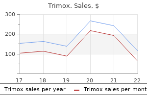
"Trimox 250mg lowest price, antibiotics quizlet".
I. Gancka, M.A., Ph.D.
Deputy Director, University of Kentucky College of Medicine
However zombie infection pc buy 500mg trimox, the early phases of the host reaction to an insect bite are often IgE-mediated or the result of the direct effects of insect venoms virus spreading in us trimox 250 mg lowest price. Important delayed-type hypersensitivity responses to divalent cations such as nickel have also been observed antibiotic treatment for mrsa purchase trimox 250 mg visa. Finally virus database generic trimox 500mg free shipping, although this section has focused on the role of T cells in inducing delayed-type hypersensitivity reactions, there is evidence that antibody and complement may also play a part. Mice deficient in B cells, antibody, or complement show impaired contact hypersensitivity reactions. These requirements for B cells, antibody, and complement may reflect their role in the early steps of the elicitation of these reactions. Hypersensitivity diseases reflect normal immune mechanisms directed against innocuous antigens. They can be mediated by IgG antibodies bound to modified cell surfaces, or by complexes of antibodies bound to poorly catabolized antigens, as occurs in serum sickness. Hypersensitivity reactions mediated by T cells can be activated by modified self proteins, or by injected proteins such as those in the mycobacterial extract tuberculin. These T cellmediated responses require the induced synthesis of effector molecules and develop more slowly, which is why they are termed delayed-type hyper-sensitivity. In some people, immune responses to otherwise innocuous antigens produce allergic or hypersensitivity reactions upon reexposure to the same antigen. Most allergies involve the production of IgE antibody to common environmental allergens. Some people are intrinsically prone to making IgE antibodies against many allergens, and such people are said to be atopic. The physiological role of this system is to provide front-line defense against parasite pathogens but, in economically developed societies in which parasitic infections are uncommon, it is almost always involved in allergic reactions. Eosinophils and specific effector T cells have an extremely important role in chronic allergic inflammation, which is the major cause of the chronic morbidity of asthma. Antibodies of other isotypes and antigen-specific effector T cells contribute to hypersensitivity to other antigens. Complexity and redundancy in the pathogenesis of asthma: reassessing the roles of mast cells and T cells J. Atopic allergy and other hypersensitivities interactions between genetic susceptibility, innocuous and/or microbial antigens and the immune system Curr. Myeloid dendritic cells induce Th2 responses to inhaled antigen, leading to eosinophilic airway inflammation J. Allergen and bacterial antigen-specific T-cell clones established from atopic donors show a different profile of cytokine production Proc. Dendritic cells with antigenpresenting capability reside in airway epithelium, lung parenchyma, and visceral pleura J. Identification of recombinant filarial proteins capable of inducing polyclonal and antigen-specific IgE and IgG4 antibodies J. Protease-dependent activation of epithelial cells by fungal allergens leads to morphologic changes and cytokine production J. House dust mite allergen characterisation: implications for T-cell responses and immunotherapy Int. Der p 1 facilitates transepithelial allergen delivery by disruption of tight junctions J. Induction of human IgE synthesis in B cells by mast cells and basophils Nature 1993. Flexible programs of chemokine receptor expression on human polarized T helper 1 and 2 lymphocytes J. The association of atopy with a gainof-function mutation in the alpha subunit of the interleukin-4 receptor N. The inverse association between tuberculin responses and atopic disorder Science 1997. Regulation of antibody responses via antibodies, complement, and Fc receptors Annu.
The spectrum of activity of the echinocandins is limited to those fungi in which 1 antimicrobial paint cheap trimox 500 mg with visa,3-glucans constitute the dominant cell wall glucan component infection lymph node safe 250mg trimox. The echinocandins have excellent activity against fluconazole-resistant strains of Candida spp antibiotics for uti septra generic trimox 500mg visa. Primary or acquired resistance to this class of agents appears to be uncommon among clinical isolates of Candida and Aspergillus species bacterial vaginosis treatment purchase 500 mg trimox with visa. The echinocandins must be administered intravenously and are highly (>95%) protein bound. They are distributed to all major organs, although concentrations in cerebrospinal fluid are low. All of the echinocandins are very well tolerated and have few drug-drug interactions. Among the three echinocandins, all have similar spectrum and potency against Candida and Aspergillus species. Echinocandins X Glucan X Caspofungin is approved for treatment of invasive candidiasis, including candidemia, and for treatment of patients with invasive aspergillosis refractory to or intolerant of other approved antifungal therapies. Anidulafungin is approved for treatment of esophageal candidiasis and candidemia, and micafungin is approved for treatment of esophageal candidiasis and candidemia and for prevention of invasive candidiasis. Flucytosine is water soluble and has excellent bioavailability when administered orally. High concentrations of flucytosine may be achieved in serum, cerebrospinal fluid, and other body fluids. Monitoring of serum concentrations of flucytosine is important in avoiding toxicity. Flucytosine is not used as monotherapy, because of the propensity for secondary resistance. Combinations of flucytosine with either amphotericin B or fluconazole have been shown to be efficacious in treating both cryptococcosis and candidiasis. Cell wall Cell membrane Allylamines the allylamine class of antifungal agents includes terbinafine, which has systemic activity, and naftifine, which is a topical agent (see Table 61-1). These agents inhibit the enzyme squalene epoxidase, which results in a decrease in ergosterol and an increase in squalene within the fungal cell membrane (see Figures 61-1 and 61-4). Terbinafine is a lipophilic antifungal agent with a broad spectrum of activity that includes dermatophytes, Candida spp. It is available in oral and topical formulations and achieves high concentrations in fatty tissues, skin, hair, and nails. Terbinafine is efficacious in the treatment of virtually all forms of dermatomycoses, including onychomycosis, and exhibits few side effects. Griseofulvin Griseofulvin is an oral agent used in the treatment of infections caused by the dermatophytes. It is thought to inhibit fungal growth by interaction with microtubules within the fungal cell, resulting in inhibition of mitosis (see Table 61-1 and Figure 61-1). Griseofulvin is considered a second-line agent in the treatment of dermatophytoses. Newer agents, such as itraconazole and terbinafine, are more rapid acting and provide greater efficacy. Griseofulvin is also associated with a number of mild side effects, including nausea, diarrhea, headache, hepatotoxicity, rash, and neurologic side effects. Inhibition of chitin synthesis in the fungal cell wall by nikkomycin Z provides another novel approach that may be useful in concert with other inhibitors of cell wall or cell membrane synthesis. The development of agents with novel mechanisms of action is both necessary and promising for future advances in the area of antifungal therapy. In addition to aggressive use of new antifungal agents such as voriconazole and caspofungin as monotherapy, the use of azole-, echinocandin-, and polyene-based combinations for treatment of the more difficult-to-treat mycoses. The rationale behind combination therapy is that by using combinations of antifungal agents, one may achieve a better clinical outcome than with monotherapy. The push toward the use of combination antifungal therapy is especially strong for infections such as invasive aspergillosis, where the associated mortality is unacceptably high. In considering combination therapy, one seeks to achieve synergy and avoid antagonism.
Among the various species of Candida capable of causing human infection (see Box 65-1 and Table 65-3) zinc vs antibiotics for acne 250 mg trimox with visa, C antibiotic resistance future trimox 250mg free shipping. Each one of these factors infection vs disease buy generic trimox 250 mg line, alone or in combination antibiotic iv trimox 250 mg generic, may affect the prevalence of different Candida spp. Likewise, breaks in infection-control precautions and in the proper care of vascular catheters may lead to more infections with C. Notably, the excess or attributable mortality resulting from candidemia has not decreased from that observed in the mid-1980s to that observed in the present day, despite the introduction of new antifungal agents with good activity against most species of Candida. Clearly, more is known about the epidemiology of nosocomial candidemia than any other fungal infection. The accumulated evidence allows one to propose a general view of nosocomial candidemia (Figure 65-4). Compared to control subjects without the specific risk factors or exposures, the likelihood of these already high-risk patients contracting candidemia in the hospital is approximately 2 times greater for each class of antibiotics they receive, 7 times greater if they have a central venous catheter, 10 times greater if Candida has been found to be colonizing other anatomic sites, and 18 times greater if they have undergone acute hemodialysis. Hospitalization in the intensive care unit setting provides the opportunity for transmission of Candida among patients and has been shown to be an additional independent risk factor. Approximately 49% of these patients will die as a result of their infection, 12% will die of their underlying disease, and 39% will survive hospitalization (see Figure 65-4). This picture has not changed (and may even be worse) from that seen in the mid-1980s. The outcome for almost half of those patients with candidemia could be improved by more effective means of prevention, diagnosis, and therapy. Clearly the most desirable of these is prevention, which is best approached by rigorous control of the exposures-especially limiting use of broad-spectrum antibiotics, improving catheter care, and adhering to infection-control practices. Infections range from superficial mucosal and cutaneous candidiasis to widespread hematogenous dissemination involving target organs such as the liver, spleen, kidney, heart, and brain. These infections are generally seen in individuals with local or generalized immunosuppression or in those settings in which candidal overgrowth is favored (see Table 65-6). Other presentations include the pseudomembranous type, which reveals a raw bleeding surface when scraped; the erythematous type-flat, red, occasionally sore areas; candidal leukoplakia-nonremovable white thickening of epithelium caused by Candida spp. These infections present as a pruritic rash with erythematous vesiculopustular lesions. Onychomycosis and paronychia may occur in the setting of a mixed microbial flora, including Candida. These lesions are of major diagnostic importance; they can be directly biopsied and thus provide an etiologic diagnosis of a systemic process. Chronic mucocutaneous candidiasis is a rare condition marked by a deficiency in T-lymphocyte responsiveness to Candida spp. These patients suffer from severe unremitting mucocutaneous Candida lesions, including extensive nail involvement and vaginitis. Benign colonization of the bladder is most common in these settings, but urethritis and/or cystitis may occur. Hematogenous seeding of the kidney may result in renal abscess, papillary necrosis, or "fungus ball" of the ureter or renal pelvis. These infections may remain localized to the abdomen, involve adjacent organs, or lead to hematogenous candidiasis. Hematogenous candidiasis may be acute or chronic and usually results in seeding of deep tissues, including the abdominal viscera, heart, eyes, bones and joints, and brain. Chronic hepatosplenic candidiasis may occur after overt or occult fungemia and presents as an indolent process marked by fever, elevated alkaline phosphatase, and multiple lesions in the liver and spleen. This process may mimic bacterial meningitis, or the course may be indolent or chronic. The clinical presentation resembles bacterial endocarditis, with fever and a new or changing heart murmur. The vegetations are classically large and friable, and embolic events are more common with endocarditis caused by Candida spp.
Overall antibiotic before surgery trimox 250mg with mastercard, the structural antimicrobial effect of aloe vera 500mg trimox free shipping, functional antibiotic for lyme disease order 500 mg trimox fast delivery, and genetic relations between the cytokines and their receptors suggest that they may have diversified in parallel during the evolution of increasingly specialized effector functions antimicrobial effects of garlic order trimox 500 mg mastercard. These specific functional effects depend on intracellular signaling events that are triggered by the cytokines binding to their specific receptors. The hematopoietin and interferon receptors all signal through a similar pathway, which is described in Chapter 6. Fas is important in maintaining lymphocyte homeostasis, as can be seen from the effects of mutations in the Fas or Fas ligand genes. Mice and humans with a mutant form of Fas develop a lymphoproliferative disease associated with severe autoimmunity. A mutation in the gene encoding the Fas ligand in another mouse strain creates a nearly identical phenotype. These mutant phenotypes represent the best-characterized examples of generalized autoimmunity caused by single-gene defects. Interactions between armed effector T cells and their targets are initiated by transient nonspecific adhesion between the cells. This recognition event triggers the armed effector T cell to adhere more strongly to the antigen-bearing target cell and to release its effector molecules directly at the target cell, leading to the activation or death of the target. The consequences of antigen recognition by an armed effector T cell are determined largely by the set of effector molecules it produces on binding a specific target cell. Membrane-associated effector molecules can deliver signals only to an interacting cell bearing the appropriate receptor, whereas soluble cytokines can act on cytokine receptors expressed locally on the target cell, or on hematopoietic cells at a distance. All viruses, and some bacteria, multiply in the cytoplasm of infected cells; indeed, the virus is a highly sophisticated parasite that has no biosynthetic or metabolic apparatus of its own and, in consequence, can replicate only inside cells. Once inside cells, these pathogens are not accessible to antibodies and can be eliminated only by the destruction or modification of the infected cells on which they depend. Physical or chemical injury, such as the deprivation of oxygen that occurs in heart muscle during a heart attack, or membrane damage with antibody and complement, leads to cell disintegration or necrosis. The dead or necrotic tissue is taken up and degraded by phagocytic cells, which eventually clear the damaged tissue and heal the wound. Apoptosis is a normal cellular response that is crucial in the tissue remodeling that occurs during development and metamorphosis in all multicellular animals. As we saw in Chapter 7, most thymocytes die an apoptotic death when they fail positive selection or, much less often, are negatively selected as a result of recognizing self antigens. The cell then destroys itself from within, shrinking by shedding membrane-bound vesicles, and degrading itself until little is left. Cytotoxic T cells kill their targets by programming them to undergo apoptosis. When cytotoxic T cells are mixed with target cells and rapidly brought into contact by centrifugation, they can program antigen-specific target cells to die within 5 minutes, although death may take hours to become fully evident. The short period required by cytotoxic T cells to program their targets to die reflects the release of preformed effector molecules, which activate an endogenous apoptotic pathway within the target cell. As well as killing the host cell, the apoptotic mechanism may also act directly on cytosolic pathogens. This prevents the assembly of virions and thus the release of infectious virus, which could otherwise infect nearby cells. Other enzymes activated in the course of apoptosis may destroy nonviral cytosolic pathogens. Apoptosis is therefore preferable to necrosis as a means of killing infected cells; in necrosis, intact pathogens are released from the dead cell and these can continue to infect healthy cells, or can parasitize the macrophages that ingest them. Each killing requires the same series of steps, including receptor binding and directed release of cytotoxic proteins stored in lytic granules. The process of apoptosis is shown in the micrographs (bottom panels), where panel a shows a healthy cell with a normal nucleus. Early in apoptosis (panel b) the chromatin becomes condensed (red) and, although the cell sheds membrane vesicles, the integrity of the cell membrane is retained, in contrast to the necrotic cell in the upper part of the same field. In late stages of apoptosis (panel c), the cell nucleus (middle cell) is very condensed, no mitochondria are visible, and the cell has lost much of its cytoplasm and membrane through the shedding of vesicles. The principal mechanism through which cytotoxic T cells act is by the calcium-dependent release of specialized lytic granules upon recognition of antigen on the surface of a target cell.
Some are useful in detecting a wide variety of parasites antimicrobial overview generic 250 mg trimox visa, and others are particularly useful for one or a few parasites infection in gums trimox 500mg fast delivery. Although the mainstay of diagnostic clinical microbiology is isolation of the causative pathogen in culture virus 0xffd12566exe buy trimox 250mg free shipping, the diagnosis of parasitic diseases is accomplished almost entirely by morphologic (usually microscopic) demonstration of parasites in clinical material bacteria quiz generic 250mg trimox fast delivery. Occasionally, demonstration of a specific antibody response (serodiagnosis) helps in establishing the diagnosis. The detection of parasite antigens in serum, urine, or stool now provides a rapid and sensitive means of diagnosing infection with certain organisms. In general, it is better for the laboratory to offer a limited number of competently performed procedures than to offer a wide variety of infrequently and poorly performed tests. This chapter provides a general description of the principles of specimen collection and processing necessary to diagnose most parasitic infections. Specific details of these and other procedures of general and limited usefulness may be found in several reference texts listed in the Bibliography. Understanding the life cycle of parasitic organisms is a key to understanding important features of geographic distribution, transmission, and pathogenesis of many parasitic diseases. For example, in the life cycle of filariae that infect humans, certain species. Sampling the blood of such patients during daytime hours may fail to detect the microfilariae, whereas blood specimens collected between 10 pm and 4 am may demonstrate many microfilariae. Likewise, intestinal nematodes such as Ascaris lumbricoides and hookworm, which reside in the lumen of the intestine, produce large numbers of eggs that can be detected easily in the stool of an infected patient. In contrast, another intestinal nematode, Strongyloides stercoralis, lays its eggs in the bowel wall rather than in the intestinal lumen. As a result, the eggs are rarely seen on stool examination; to make the diagnosis, the parasitologist must be alert for the presence of larvae. Finally, parasites may cause clinical symptoms at a time when diagnostic forms are not yet present in the usual site. For example, in certain intestinal nematode infections, migration of larvae through the tissues may cause intense symptomatology weeks before the characteristic eggs are present in feces. Because the majority of parasitologic examinations and identifications are based entirely on recognizing the characteristic morphology of the organisms, any condition that may obscure or distort the morphologic appearance of the parasite may result in an erroneous identification or missed diagnosis. As noted previously and in Box 71-1, there may be alternatives to microscopy for detection and identification of certain parasites. Sampling of perianal skin is a useful means of recovering the eggs of Enterobius vermicularis (pinworm) or Taenia species (tapeworm). Fecal Specimen Collection Patients, clinicians, and laboratory personnel must be properly instructed on collection and handling of specimens. Fecal specimens should be collected in clean wide-mouthed, waterproof containers with a tight-fitting lid to ensure and maintain adequate moisture. Specimens must not be contaminated with water, soil, or urine, because water and soil may contain free-living organisms that can be mistaken for human parasites, and urine can destroy motile trophozoites and may cause helminth eggs to hatch. Stool specimens should not contain barium, bismuth, or medications containing mineral oil, antibiotics, antimalarials, or other chemical substances, because such specimens compromise the detection of intestinal parasites. Specimen collection should be delayed for 5 to 10 days to allow barium to clear and for at least 2 weeks after antibiotics such as tetracycline to allow intestinal parasites to recover from the toxic (but not curative) effects of the drugs. Purged specimens may be collected when organisms are not detected in normally passed fecal specimens; however, only certain purgatives (sodium sulfate and buffered sodium biphosphate [phosphosoda]) are satisfactory. One series of purged specimens may be examined in place of or in addition to a series of normally passed specimens. Unpreserved formed fecal specimens should arrive in the laboratory within 2 hours after passage. If the stool is liquid and thus more likely to contain trophozoites, it should reach the laboratory for examination within 30 minutes. The number of specimens required to demonstrate intestinal parasites varies depending on the quality of the specimen submitted, the accuracy of the examination performed, the severity of the infection, and the purpose for which the examination is made. If the physician is interested only in determining the presence or absence of helminths, one or two examinations may suffice, provided concentration methods are used. For a routine parasitic examination, a total of three fecal specimens is recommended.
