A distinctive characteristic of these blasts is the linear purple Auer rod composed of crystallized azurophilic granules gastritis gi bleed generic rabeprazole 10 mg on-line. The M2 kind seen right here lymphocytic gastritis definition rabeprazole 20 mg cheap mastercard, the commonest, has distinguished Auer rods with a range of immature to mature myeloid cells present. Thus this marrow is basically 100% cellular, but with leukemic blasts almost exclusively filling the marrow-more than the 20% or more blasts wanted for diagnosis-displacing and changing regular hematopoiesis (a myelophthisic process) or suppressing stem cell division. Leukemic patients are prone to anemia, thrombocytopenia, and granulocytopenia, and all of the issues that ensue, significantly complications of bleeding and infection. For this purpose, remedy with retinoic acid, a vitamin A analogue, helps overcome the block. Cell demise with release of the granules into the peripheral blood may cause disseminated intravascular coagulation. Bone marrow findings embrace dyserythropoietic modifications with nuclear abnormalities, ringed sideroblasts in erythroid precursors (shown here with iron stain), hypogranulation and hyposegmentation in myeloid precursors, elevated myeloblasts, and lowered numbers of disorganized nuclei inside megakaryocytes. Because some instances come up from malignant transformation in a pluripotent cell line, there may be erythroid and megakaryocytic involvement. They typically terminate in marrow fibrosis, with pancytopenia, extramedullary hematopoiesis, and splenomegaly. Although congestive splenomegaly is unlikely to exceed 1000 g, a spleen larger than 1000 g suggests underlying myeloproliferative, lymphoproliferative, or hematopoietic problems. Chronic infections corresponding to malaria or leishmaniasis also might produce marked splenomegaly. Peripheral blood findings in myeloproliferative problems include leukoerythroblastosis and large platelets. A unifocal to multifocal kind known as eosinophilic granuloma primarily entails bones in kids and young adults. A multilocular eosinophilic granuloma of bone is seen here in the best higher femur. The characteristic cell is an oval to spherical, macrophage-like cell, interspersed with inflammatory cells, including eosinophils. This lesion varieties within the marrow cavity and may expand to trigger erosion of bone, which produces pain or pathologic fracture. Some lesions heal spontaneously by fibrosis, whereas others may require curettage. There is also an uncommon condition generally known as splenosis that occurs when portions of a disrupted spleen (usually from blunt belly trauma) implant on peritoneal surfaces and grow and continue to perform, even after the damaged spleen has been eliminated ("born again" spleen). The spleen also acts as a storage space for about one third of circulating platelets. Abnormal macromolecules produced with some inborn errors of metabolism, such as Gaucher disease and Niemann-Pick illness, may accumulate in splenic phagocytes and lead to splenomegaly. The spleen, a key a half of the immune system, has dendritic cells in periarterial lymphatic sheaths that lure antigens and present them to T lymphocytes, the place T and B cells work together at the edges of white pulp follicles, generating antibody-secreting plasma cells found primarily inside the sinuses of purple pulp. The lack of splenic perform from splenectomy or with autoinfarction (sickle cell disease) leads to susceptibility to disseminated infection with encapsulated bacteria, such as pneumococcus, meningococcus, and Haemophilus influenzae. The "barrel ribs" of reticulin fibers define longitudinal sinusoids within the red pulp by way of which the blood flows. Micronodular cirrhosis from persistent alcohol abuse or macronodular cirrhosis after hepatitis B or C an infection could result in portal hypertension. This spleen additionally shows irregular tan-white fibrous plaques over the purple capsular floor. The increased portal venous stress causes dilation of sinusoids, with slowing of blood move from the cords to the sinusoids that prolongs the exposure of the blood cells to the cordal macrophages, resulting in excessive trapping and destruction (hypersplenism). Portions of the friable vegetations have embolized to the spleen via the splenic artery branch from the celiac axis after which to the peripheral splenic artery branches. Most splenic infarcts are ischemic and attributable to emboli from either vegetations on valves or mural thrombi within the heart. The remaining splenic parenchyma of darkish purple pulp has pinpoint pale malpighian corpuscles of white pulp. Shown listed here are two giant capsular lacerations in a patient who was concerned in a motorcar collision. Note the darkish pink hematoma formation at the left ensuing from the splenic rupture. Enlargement of the spleen from predisposing circumstances that render the spleen susceptible to rupture even with minor trauma embrace infectious mononucleosis, malaria, typhoid fever, and lymphoid neoplasms. The peritoneal lavage performed on this affected person yielded bloody fluid, a clue to the analysis. The spleen might have to be surgically removed after such damage with splenic capsular rupture as a result of the capsule is difficult to repair. Splenic preservation is desirable to preserve immune function, significantly in youngsters. This younger patient had an inborn error of metabolism with lack of the enzyme glucocerebrosidase, leading to accumulation of storage product in cells of the mononuclear phagocyte system. The most typical, type 1 (99% of patients), is the non-neurologic kind by which the affected person has normal intelligence and lives into maturity. Numerous clusters of these macrophages enlarge the spleen, an appearance typical of a storage illness. This accumulation in marrow might produce a mass impact with bone ache, deformity, and fracture; pancytopenia can occur. Other tissues of the mononuclear phagocyte system, including lymphoid tissues and liver, can also be concerned. Perivascular accumulation of macrophages in sort 2 Gaucher disease leads to neuronal loss. As in many genetic illnesses resulting from enzymatic abnormalities, enzyme activity, on this case glucocerebrosidase exercise, in peripheral blood leukocytes or skin fibroblasts may be measured to affirm the analysis. Different alleles may result in completely different enzymatic activities, with variable severity of the illness. An immunodeficiency state leads to reduced cell-mediated immunity to battle the mycobacteria. There is eventual atrophy with fatty substitute, shown with the smaller adult thymus in the right panel. In improvement of the immune system, the thymus is a vital place to which marrow stem cells migrate, undergo maturation with selection, and provides rise to T lymphocytes. Hassall corpuscles composed of epithelial cells are in the center of the medullary areas. In embryonic improvement of the immune system, progenitor cells of marrow origin migrate to the thymus and provides rise to mature T cells that are exported to the periphery. The thymic production of T cells slowly declines throughout maturity as the organ atrophies. In addition to the thymocytes and epithelial cells, macrophages, dendritic cells, few B lymphocytes, rare neutrophils and eosinophils, and scattered myoid (muscle-like) cells are discovered within the thymus. Ordinarily the thymus in an grownup is composed principally of adipose tissue, with a number of clusters of lymphocytes and residual Hassall corpuscles. The acetylcholine receptor antibodies diminish the receptor operate within the skeletal muscle motor finish plates, resulting in the onset of muscular weak spot, significantly with repetitive muscular contraction. Most are discovered in the anterosuperior mediastinum, but sometimes they occur in the neck, thyroid, pulmonary hilus, or elsewhere. It is arising within the left side of the thymus anterior and to the left of the aortic arch. Thymomas could be gradual rising and act in a benign fashion, but malignant thymomas could be locally invasive. The remainder are found by the way throughout imaging research or during cardiothoracic surgery. The neoplastic epithelial elements of this thymoma display minimal pleomorphism, however this tumor was regionally invasive and a malignant thymoma. The cells could be marked and enumerated to assist classify them into a diagnostic category. A myeloproliferative course of can contain the marrow, filling it up with abnormal mobile proliferations. The continual myeloproliferative process might continue for years or could blast out right into a leukemia or burn out right into a myelofibrosis. This is amyloidosis of the spleen, which might have either the diffuse lardaceous sample seen here, or the nodular sago sample with amyloid deposited mainly within the white pulp.
The focal lesions sometimes start within the medullary cavity gastritis diet áèòâà rabeprazole 10 mg discount without a prescription, erode cancellous bone gastritis garlic buy rabeprazole 20 mg low cost, and progressively destroy the bony cortex, leading to pathologic fractures, sometimes vertebral compressed fractures. The bone lesions appear radiographically as "punched-out" defects, normally 1 to four cm in diameter. About 3% to 5% of plasma cell neoplasms are solitary, however many progress to myeloma. Myelomas are typically detected by an immunoglobulin spike on protein electrophoresis or by the presence of Bence Jones proteins (light chains) within the urine. Immunoelectrophoresis characterizes the kind of monoclonal immunoglobulin being produced. There are numerous well-differentiated plasma cells with eccentric nuclei and a perinuclear halo of clearer cytoplasm (representing the Golgi apparatus). This neoplasm is typically well differentiated, with easily recognizable plasma cells, most of that are hardly distinguishable from normal plasma cells except by their increased numbers. It has plasmacytoid differentiation in order that many cells resemble plasma cells and may secrete immunoglobulin, usually as an IgM paraproteinemia. Many of these neoplasms secrete substantial quantities of monoclonal IgM, with Waldenstr�m macroglobulinemia, resulting in a hyperviscosity syndrome with scientific findings that embrace decreased vision, complications, dizziness, coagulopathy, and cryoglobulinemia with cold agglutinins and hemolytic anemia. The attribute karyotypic abnormality is t(11;14) with fusion of the cyclin D1 gene on chromosome 11 to the immunoglobulin heavychain promoter-enhancer area on chromosome 14, leading to increased cyclin D1 expression with loss of cell cycle regulation. The preliminary polyclonal enlargement might acquire t(11;18) or t(14;18) and evolve to monoclonal lymphoma. The spleen has uniform attenuation, typical for diffuse lymphoid neoplasms, which are often diffusely infiltrative and infrequently necrotic or hemorrhagic. It usually manifests with extranodal multiorgan involvement (skin, bone marrow, delicate tissues, liver) along with fever. Note the small cells with convoluted nuclei infiltrating the dermis (left panel) and increasing into epidermis as Pautrier microabscesses (right panel). An inflammatory premycotic phase progresses by way of a plaque part to a tumor phase on the pores and skin. S�zary syndrome happens when pores and skin involvement is manifested by generalized exfoliative erythroderma and an related leukemia of S�zary cells, also referred to as S�zary-Lutzner cells, with characteristic cerebriform nuclei. Note the appearance of the deep-clefted, cerebriform nucleus on this circulating lymphocyte. Late in the course of this illness, transformation to a big T-cell lymphoma typically happens. It is essential to decide whether or not the patient has solely a single lymph node area involved, a number of node areas, or extranodal involvement. Many sufferers respond to chemotherapy, notably youthful patients, sufferers with a decrease stage of illness, and people with absence of constitutional symptoms. The giant multinucleated cells proven on this effaced lymph node typically constitute solely 1% to 5% of the cellular mass of the neoplasm, with the remainder composed of reactive cells and connective tissue. Clinical findings in about 40% of sufferers may embody constitutional ("B") symptoms, such as fever, evening sweats, and weight loss. They secrete cytokines that promote proliferation of extra reactive cells, forming the majority of the tumor mass. Cell-mediated immunity is commonly decreased, as evidenced by anergy with skin testing and more frequent and extreme infections. Laboratory findings embody anemia, leukocytosis, and an elevated erythrocyte sedimentation price. The background infiltrate of cells has lymphocytes, plasma cells, eosinophils, and macrophages. Compared with the lymphocyte predominance and nodular sclerosis subtypes, combined cellularity subtype is extra more doubtless to be related to older age, systemic symptoms such as night time sweats, and weight loss. Advanced stage and systemic signs are frequent, and the overall outcome is much less favorable than with other subtypes. In most circumstances, lymph nodes are diffusely effaced, however obscure nodularity resulting from the presence of residual B-cell follicles can generally be seen. The lymphocyte rich variant has frequent mononuclear variants and Reed-Sternberg cells. This peripheral blood smear exhibits giant, immature myeloblasts with nuclei which have fantastic chromatin and a number of nucleoli. Diffuse enlargement with out focal lots could result from leukemic involvement or extramedullary hematopoiesis. Most of these masses are tan, but some have brown-black pigmentation from the melanin elaborated by the neoplastic cells. Spindle-cell thymomas are often well-circumscribed neoplasms that grow slowly, with an indolent course. Such cells wish to travel and assist to collect and transfer antigens to the lymphocytes for recognition and immune activation. This marrow was taken from the posterior iliac crest of a middle-aged particular person; marrow is about 50% mobile at age 50 years, declining by 10% per decade thereafter. In very elderly people, most remaining hematopoiesis is concentrated in vertebrae, sternum, and ribs. The erythroid islands and granulocytic precursors type the majority of the hematopoietic parts, admixed with steatocytes. Small numbers of lymphocytes, primarily reminiscence B cells, and plasma cells secreting immunoglobulins are present. A normal mature lymphocyte on the left can be compared with a segmented neutrophil (polymorphonuclear leukocyte) on the best. This phenomenon happens with a rise in serum proteins, particularly fibrinogen, C-reactive protein, and globulins. The bone marrow can respond to anemia with elevated erythropoiesis, indicated by an elevated reticulocyte count. This lack produces membrane instability that forces the cell to take the smallest volume possible-a sphere. This X-linked dysfunction, present in 12% of male African Americans, can additionally be seen in people from the Mediterranean region, including Italy, Greece, and Turkey. It is asymptomatic until stress occurs from infection or ingestion of an oxidizing drug. Laboratory findings embody anemia, reticulocytosis, indirect hyperbilirubinemia, and decreased haptoglobin. Shown here in lane 1 is an infant with sickle cell anemia (hemoglobin S [Hgb S]) and vital Hgb F manufacturing; the heterozygous state of sickle cell trait is proven in lane 4. Hemoglobin electrophoresis in sickle cell disease shows 90% to 95% Hgb S, 1% to 3% Hgb A2, and 5% to 10% Hgb F. Lack of a spleen predisposes to infections, particularly with encapsulated bacterial organisms corresponding to pneumococcus. In African Americans, the gene frequency for Hgb S is about 1 in 25, with a provider fee of 1 in 12, and a 1 in 625 probability for sickle cell illness. The irregular -globin gene has an amino acid substitution at position 6 (6Glu-Lys). The resulting anemia tends to be delicate, even with homozygous illness proven in the best panel. The abnormal -globin gene has an amino acid substitution at place 26, with lysine substituted for glutamic acid. This affected person has -thalassemia, a hereditary disorder with poor -globin chain synthesis that results in ineffective erythropoiesis and a continual, microcytic anemia. There is inadequate manufacturing of Hgb F and A2 to fully compensate for reduced A1. The residual function and -globin manufacturing determine the severity of the anemia. Such an increase in marrow in skull could result in "frontal bossing" or forehead prominence due to the skull form change. The outcome can be bony prominence with prominent epiphyseal regions, as seen on this 20-year-old man. The hemochromatosis can result in dilated cardiomyopathy, hepatic failure, hypogonadism, and "bronze" diabetes mellitus from iron deposition in islets of Langerhans. The tetramer of chains types Hgb H, which may be seen in small amounts in neonates with -Thalassemia minor. Larger quantities (10% to 25%) of Hgb H persist in persons with three irregular -globin chains (Hgb H disease). Anisocytosis (variation in size) and poikilocytosis (variation in shape) are additionally increased.
It can additionally be associated to quite a few other structures: Anterior to the belly aorta gastritis symptoms back pain quality rabeprazole 10 mg, because it descends gastritis diet 911 order 20 mg rabeprazole otc, are the pancreas and splenic vein, the left renal vein, and the inferior a half of the duodenum. On its right side are the cisterna chyli, thoracic duct, azygos vein, right crus of the diaphragm, and the inferior vena cava. Vasculature Abdominal aorta the stomach aorta begins at the aortic hiatus of the diaphragm as a midline construction at approximately the Inferior phrenic arteries Diaphragm Celiac trunk Middle s uprarenal artery Middle s uprarenal artery Left renal artery Superior mes enteric artery Tes ticular or ovarian arteries Lumbar arteries Inferior mes enteric artery Common iliac artery Ps oas major mus cle Median s acral artery 196. The three unpaired visceral branches that arise from the anterior surface of the abdominal aorta (Table four. They run laterally and posteriorly over the bodies of the lumbar vertebrae, continue laterally, passing posterior to the sympathetic trunks and between the transverse processes of adjoining lumbar vertebrae, and attain the stomach wall. From this level onward, they show a branching pattern much like a posterior intercostal artery, which incorporates offering segmental branches that offer the spinal wire. This vessel arises from the posterior floor of the stomach aorta simply superior to the bifurcation and passes in an inferior path, rst over the anterior floor of the lower lumbar vertebrae after which over the anterior floor of the sacrum and coccyx. Clinical app Abdominal aortic stent graft An belly aortic aneurysm is a dilatation of the aorta and generally tends to occur in the infrarenal region (the area at or under the renal arteries). Treatment of aneurysms previous to rupture can involve inserting an endovascular graft. The method includes surgically dissecting the femoral artery under the inguinal ligament. A small incision is made in the femoral artery and the preloaded compressed graft with steel help struts is passed on a big catheter into the belly aorta through the femoral artery. Using X-ray for guidance the graft is opened in order that it traces the within of the aorta. Posterior branches the posterior branches of the belly aorta are vessels supplying the diaphragm or physique wall and are (Table four. Whatever their origin, they pass upward, present some arterial supply to the suprarenal gland, and proceed onto the inferior floor of the diaphragm. Tributaries to the inferior vena cava embody the: frequent iliac veins, lumbar veins, proper testicular or ovarian vein, renal veins, right suprarenal vein, inferior phrenic veins, and hepatic veins. Of the venous tributaries talked about above, the lumbar veins are unique of their connections and deserve particular consideration. Not all of the lumbar veins drain directly into the inferior vena cava: the fth lumbar vein generally drains into the iliolumbar vein, a tributary of the common iliac vein. Note the photographs solely demonstrate the intraluminal contrast and not the whole vessel. Hepatic veins Es ophagus Inferior vena cava Left kidney Left renal vein Right tes ticular or ovarian vein Right kidney Inferior vena cava the inferior vena cava returns blood from all structures beneath the diaphragm to the best atrium of the center. It ascends via the posterior belly area anterior to the vertebral column immediately to the right of the stomach aorta. During its course, the anterior floor of the inferior vena cava is crossed by the proper common iliac artery, the root of the mesentery, the best testicular or ovarian Abdominal aorta Right external iliac artery and vein Left exterior iliac artery and vein Left femoral artery and vein Right femoral artery and vein 198. Regional anatomy � Posterior abdominal area the ascending lumbar veins are lengthy, anastomosing venous channels that connect the widespread iliac, iliolumbar, and lumbar veins with the azygos and hemiazygos veins of the thorax. If the inferior vena cava becomes blocked, the ascending lumbar veins turn out to be necessary collateral channels between the lower and higher parts of the physique. Common predisposing factors embody hospitalization, surgery, oral contraceptives, smoking, and air travel. The analysis of deep vein thrombosis may be dif cult to establish, with symptoms together with leg swelling and ache and discomfort within the calf. Occasionally the clot might dislodge and move into the venous system by way of the best aspect of the center and into the principle pulmonary arteries. If the clots are of signi cant dimension they hinder blood ow to the lung and may produce instantaneous death. Other issues include destruction of the traditional valvular system in the legs, which can result in venous incompetency and continual leg swelling with ulceration. Surface anatomy Visualizing the position of main blood vessels Each of the vertebral ranges within the abdomen is related to the origin of main blood vessels. Lymphatic system Lymphatic drainage from most deep constructions and regions of the physique below the diaphragm converges mainly on collections of lymph nodes and vessels related to the major blood vessels of the posterior abdominal region. Pre-aortic and lateral aortic or lumbar nodes (para-aortic nodes) Approaching the aortic bifurcation, the collections of lymphatics associated with the 2 widespread iliac arteries and veins merge, and multiple groups of lymphatic vessels and nodes associated with the abdominal aorta and inferior vena cava cross superiorly. These collections could additionally be subdivided into pre-aortic nodes, which are anterior to the stomach aorta, and proper and left lateral aortic or lumbar nodes (para-aortic nodes), which are positioned on either facet of the abdominal aorta (Table 4. The lateral aortic or lumbar lymph nodes (para-aortic nodes) receive lymphatics from the body wall, the kidneys, the suprarenal glands, and the testes or ovaries. The pre-aortic nodes are organized across the three anterior branches of the belly aorta that offer the stomach part of the gastrointestinal tract, as well as the spleen, pancreas, gallbladder, and liver. They are divided into celiac, superior mesenteric, and inferior mesenteric nodes, and receive lymph from the organs supplied by the equally named arteries. Finally, the lateral aortic or lumbar nodes type the proper and left lumbar trunks, whereas the pre-aortic nodes form the intestinal trunk. These trunks come collectively and form a con uence that, at instances, seems as a saccular dilation (the cisterna chyli). Lymphatic vessel Right jugular trunk Left jugular trunk Right subclavian trunk Left subclavian trunk Right bronchomediastinal trunk Left bronchomediastinal trunk Area drained Right facet of head and neck Left side of head and neck Right upper limb, tremendous cial areas of thoracic and upper abdominal wall Left higher limb, tremendous cial areas of thoracic and upper stomach wall Right lung and bronchi, mediastinal structures, thoracic wall Left lung and bronchi, mediastinal structures, thoracic wall Lower limbs, stomach walls and viscera, pelvic walls and viscera, thoracic wall 200 Thoracic duct Regional anatomy � Posterior belly region four Clinical app Retroperitoneal lymph node surgical procedure From a medical perspective, retroperitoneal lymph nodes are arranged in two groups. The pre-aortic lymph node group drains lymph from the embryological midline constructions, such because the liver, bowel, and pancreas. The para-aortic lymph node group (the lateral aortic or lumbar nodes), on both side of the aorta, drain lymph from bilateral buildings, such because the kidneys and adrenal glands. Organs embryologically derived from the posterior stomach wall also drain lymph to these nodes. Massively enlarged lymph nodes are a function of lymphoma, and smaller lymph node enlargement is observed within the presence of infection and metastatic malignant spread of disease. The surgical strategy to retroperitoneal lymph node resection involves a lateral paramedian incision in the midclavicular line. The three layers of the anterolateral stomach wall (external oblique, inner indirect, and transversus abdominis) are opened and the transversalis fascia is divided. Instead of coming into the parietal peritoneum, which is commonplace process for most intra-abdominal surgical operations, the surgeon gently pushes the parietal peritoneum towards the midline, which moves the intra-abdominal contents and allows a transparent view of the retroperitoneal constructions. On the left, the para-aortic lymph node group (lateral aortic or lumbar nodes) are easily demonstrated with a clear view of the abdominal aorta and kidney. On the proper the inferior vena cava is demonstrated, which has to be retracted to entry to the right para-aortic lymph node chain (lateral aortic or lumbar nodes). The process of the retroperitoneal lymph node dissection is extremely properly tolerated and lacks the issues of entering the peritoneal cavity. Unfortunately, the complication of a vertical incision within the midclavicular line is to divide the segmental nerve provide to the rectus abdominis muscle. This produces muscle atrophy and asymmetrical proportions to the anterior stomach wall. Sympathetic trunks and splanchnic nerves the sympathetic trunks move by way of the posterior belly region anterolateral to the lumbar vertebral bodies, before continuing throughout the sacral promontory and into the pelvic cavity. These characterize collections of neuronal cell bodies-primarily postganglionic neuronal cell bodies-which are located exterior the central nervous system. There are usually four ganglia alongside the sympathetic trunks in the posterior stomach (lumbar) region. Also associated with the sympathetic trunks in the posterior abdominal region are the lumbar splanchnic nerves. These components of the nervous system pass from the sympathetic trunks to the plexus of nerves and ganglia related to the belly aorta. Usually two to four lumbar splanchnic nerves carry preganglionic sympathetic bers and visceral afferent bers. Abdominal prevertebral plexus and ganglia the abdominal prevertebral plexus is a community of nerve bers surrounding the stomach aorta. It extends from the aortic hiatus of the diaphragm to the bifurcation of the aorta into the right and left widespread iliac arteries. Continuing inferiorly, the plexus of nerve bers extending from slightly below the superior mesenteric artery to the aortic bifurcation is the belly aortic plexus. At the bifurcation of the abdominal aorta, the stomach prevertebral plexus continues inferiorly as the superior hypogastric plexus.
Fc receptors for different Ig heavy chain isotypes are expressed on many leukocyte populations and serve numerous capabilities in immunity juice diet gastritis rabeprazole 20 mg cheap with mastercard. Of these Fc receptors gastritis medical definition rabeprazole 20 mg order with visa, the ones which are most important for phagocytosis of opsonized particles are receptors for the heavy chains of IgG antibodies, known as Fc receptors, and these are the receptors that shall be primarily thought-about in this chapter. In Chapter 5, we described the neonatal Fc receptor (FcRn), which is expressed in the placenta, and on vascular endothelium and different cell types and has unique capabilities related to IgG transport across the placenta and the safety of antibodies of this isotype from turnover. Fc receptors have been classified into three teams, based on their affinities for heavy chains of different IgG subclasses. IgG4 has a really low affinity for activating Fc receptors, and the biological operate of this antibody isotype is poorly understood. All Fc receptors comprise a ligand-binding chain, referred to as the chain, that recognizes IgG heavy chains. Differences in specificities or affinities of every FcR for the various IgG isotypes are based mostly on variations in the structure of those chains. All Fc receptors are optimally activated by antibodies sure to their antigens and not by free, circulating antibodies. There are three main teams of IgG-specific Fc receptors, of which two have multiple isoforms that differ in structure and performance (see Table 13. The FcRn has unique capabilities associated to IgG transport across the placenta and the protection of antibodies of this isotype from turnover, as discussed in Chapter 5. It is expressed on macrophages and neutrophils and binds IgG1 and IgG3 with excessive affinity (dissociation constant [Kd] of 10-8 to 10-9 M). Schematic fashions of the completely different human Fc receptors illustrate the Fc-binding chains and the signaling subunits. Cross-linking of several Fc receptorbound IgG molecules by multivalent antigens ends in cell activation. These isoforms have comparable extracellular domains and ligand specificities but differ in cytoplasmic tail construction, cell distribution, and functions. On dendritic cells, this receptor can contribute to antigen capture and consequently T cell activation. In addition to these Fc receptors, there are receptors for the heavy chains of IgE and IgA (see Table 13. Role of Fc Receptors in Phagocytosis and Activation of Phagocytes Binding of Fc receptors on phagocytes to multivalent antibody-coated particles results in engulfment of the particles and activation of the phagocytes. The IgG subtypes that bind greatest to these receptors (IgG1 and IgG3) are the most environment friendly opsonins for selling phagocytosis. Opsonized particles are internalized into vesicles generally known as phagosomes, which fuse with lysosomes, and the phagocytosed particles are destroyed in these phagolysosomes. Cross-linking of the ligand-binding chains of an FcR leads to signal transduction occasions which are just like those who occur after antigen receptor cross-linking in lymphocytes (see Chapter 7). Antibodies of certain IgG subclasses bind to microbes and are then acknowledged by Fc receptors on phagocytes. Signals from the Fc receptors promote the phagocytosis of the opsonized microbes and activate the phagocytes to destroy these microbes. These occasions result in era of inositol trisphosphate and diacylglycerol and sustained increase in cytosolic calcium. The signaling pathways downstream of Fc receptors induce numerous responses in leukocytes, including transcription of genes encoding cytokines, inflammatory mediators and microbicidal enzymes, and mobilization of the cytoskeleton resulting in phagocytosis, granule exocytosis, and cell migration. The main microbicidal substances produced in the activated phagocytes are reactive oxygen species, nitric oxide, and hydrolytic enzymes. These are the identical substances produced by phagocytes activated in innate immune responses, discussed in Chapter 4. The identical microbicidal substances could damage tissues; this mechanism of antibody-mediated tissue damage is necessary in hypersensitivity ailments (see Chapter 19). Antibody-Mediated Clearance of Helminths Antibodies, eosinophils, and mast cells operate collectively to mediate the killing and expulsion of some helminthic parasites. Helminths (worms) are too large to be engulfed by phagocytes, and their integuments are relatively proof against the microbicidal merchandise of neutrophils and macrophages. They can, nonetheless, be killed by a poisonous cationic protein, known as the most important primary protein, present within the granules of eosinophils. IgE and, to a lesser extent, IgG and IgA antibodies that coat helminths can bind to Fc receptors on eosinophils and trigger the degranulation of these cells, releasing the essential protein and different eosinophil granule contents that kill the parasites. In addition to activating eosinophils, IgE antibodies that acknowledge antigens on the floor of the helminths might provoke local mast cell degranulation by way of the high-affinity IgE receptor (see Chapter 20). Mast cell mediators may induce bronchoconstriction and increased intestinal motility, contributing to the expulsion of worms from sites such as the airways and the lumen of the gastrointestinal tract. The name complement is derived from experiments performed by Jules Bordet shortly after the invention of 282 Chapter thirteen � Effector Mechanisms of Humoral Immunity antibodies. He demonstrated that if recent serum containing an antibacterial antibody is added to the bacteria at physiologic temperature (37�C), the micro organism are lysed. Bordet concluded that the serum must contain one other heat-labile component that assists, or enhances, the lytic operate of antibodies, and this component was later given the name complement. The complement system consists of serum and cell surface proteins that work together with each other and with other molecules of the immune system in a highly regulated method to generate products that perform to get rid of microbes. Pathways of Complement Activation There are three major pathways of complement activation: the classical pathway, which is activated by certain isotypes of antibodies bound to antigens; the alternative pathway, which is activated on microbial cell surfaces in the absence of antibody; and the lectin pathway, which is activated by mannose-binding protein that binds to floor carbohydrates on microbes. The names classical and alternative arose as a end result of the classical pathway was found and characterized first, but the various pathway is phylogenetically older. The alternative and lectin pathways are effector mechanisms of innate immunity, whereas the classical pathway is a significant mechanism of adaptive humoral immunity. The central occasion in complement activation is proteolysis of the complement protein C3 to generate biologically active products and the following covalent attachment of a product of C3, known as C3b, to microbial cell surfaces or to antibody certain to antigen. Complement activation entails the generation of a proteolytic complex, the C3 convertase, which cleaves C3 into two fragments referred to as C3a and C3b. All of the biologic functions of complement are depending on the proteolytic cleavage of C3. For example, complement activation promotes phagocytosis as a result of C3b turns into covalently linked to microbes, and phagocytes (neutrophils and macrophages) categorical receptors for C3b. Peptides produced by proteolysis of C3 (and other complement proteins) stimulate inflammation. In all three pathways of complement activation, after the era of C3b by the C3 convertase, a second enzyme complex known as the C5 convertase is assembled, which cleaves C5 into C5a and C5b. The C5 convertase contributes both to inflammation by era of the C5a fragment, and to the formation of pores within the membranes of microbial targets. The pathways of complement activation differ in how C3b is produced however follow a typical sequence of reactions after the cleavage of C5. With this background, we proceed to more detailed descriptions of the choice, classical, and lectin pathways. The Alternative Pathway the choice pathway of complement activation results in the proteolysis of C3 and the secure attachment of its breakdown product C3b to microbial surfaces, with no role for antibody. Normally, C3 in plasma is being repeatedly cleaved at a low price (1% to 2% of the total plasma C3 per hour) to generate C3b in a process that is identified as C3 tickover. Activation of complement involves the sequential proteolysis of proteins to generate enzyme complexes with proteolytic activity. Proteins that purchase proteolytic enzymatic activity by the motion of other proteases are called zymogens. The means of sequential zymogen activation, a defining characteristic of a proteolytic enzyme cascade, can be characteristic of the coagulation and kinin systems. Proteolytic cascades enable large and speedy amplification as a end result of every enzyme molecule activated at one step can generate multiple activated enzyme molecules at the next step. Many of the biologically active cleavage products of complement activation become covalently attached to microbial cell surfaces, to antibodies certain to microbes and different antigens, and to apoptotic our bodies. Recruitment of neutrophils and monocytes establishes an inflammatory environment round microbes that helps to eliminate the pathogens. Complement activation is inhibited by regulatory proteins which may be present on normal host cells and absent from microbes. The regulatory proteins are an adaptation of normal cells that decrease complementmediated harm to host cells. Because microbes lack these regulatory proteins, complement activation can happen on microbial surfaces. The various pathway is activated by C3b binding to numerous activating surfaces, similar to microbial cell partitions; the classical pathway is initiated by C1 binding to antigen-antibody complexes; and the lectin pathway is activated by binding of a plasma lectin to microbes. The late steps of all three pathways are the identical (not shown), and complement activated by all three pathways serves the same capabilities.
Shown right here is an embolic infarct involving a cerebral hemisphere in the left panel gastritis kronik generic rabeprazole 10 mg overnight delivery, which microscopically exhibits features of a mycotic aneurysm in the proper panel chronic gastritis outcome buy rabeprazole 20 mg low cost, with destruction of an arterial wall by the blue bacterial colonies. The main benefit of this bioprosthesis is the dearth of need for continued anticoagulation. The drawback of this kind of prosthetic coronary heart valve is the restricted life span of the prosthetic cusps, on common 5 to 10 years (but generally shorter), because of wear and subsequent dystrophic calcification that reduces cusp motion and results in stenosis. Such mechanical prostheses final indefinitely from a structural standpoint, however the affected person requires continuing anticoagulation because the uncovered nonbiologic surfaces are susceptible to thrombosis. The inferior facet is proven in the left panel with the left ventricular chamber below. The outflow tract from this prosthesis is shown in the best panel, with the 2 leaflets tilted outward towards the left atrium. Another prosthetic complication is infective endocarditis, which is most vulnerable to involve the ring. Although these mechanical prostheses final indefinitely from a structural standpoint, the affected person requires continuing anticoagulation to prevent thrombosis. The valve cusps are nonetheless pliable, however the valve has turn out to be contaminated with giant vegetation filling the valve orifice. The leads from the battery prolong all the way down to the best atrium and the apex of the right ventricle. It felt very flabby at autopsy, and the myocardium in life was poorly contractile. Some circumstances may occur after myocarditis, whereas others may appear within the peripartum period with pregnancy. This identical appearance may comply with ischemic damage, by which case the term ischemic cardiomyopathy might be applied. In most circumstances of idiopathic dilated cardiomyopathy, the coronary arteries show little or no atherosclerosis. It may be idiopathic or the result of an identifiable cause, such as hemochromatosis, with excessive iron deposition, proven right here with Prussian blue iron stain. The iron deposition leads to myocardial dysfunction with cardiac coronary heart enlargement and failure. Iron overload can accompany hereditary hemochromatosis or transfusion-dependent anemias. This condition has an incidence of 1 in 10,000, and could additionally be familial, with an autosomal dominant pattern of transmission with mutations in genes encoding for desmosomal proteins such as plakoglobin affecting hole junctions. This condition is related to dysrhythmias that may result in syncope and even sudden dying from heart failure. The pathogenesis of this situation includes irregular desmosome proteins with uncontrolled apoptosis of myocardium, muscular degeneration with fibrofatty replacement, and inflammation. Note the marked left ventricular hypertrophy and asymmetric bulging of a really large interventricular septum into the left ventricular chamber. One in 500 persons is affected, and about half of circumstances are familial, although various gene mutations may be answerable for this illness. Children and adults could be affected, and sudden dying can occur, sometimes from an arrhythmia. In the left panel with H&E stain and in the best panel with trichrome stain are sections of myocardium showing these irregular myofibers with surrounding collagen. Many circumstances are caused by mutations in genes encoding for sarcomeric proteins, such as myosin heavy chain, troponin T, myosin-binding protein C, and -tropomyosin. Clinical findings are associated to decreased ventricular compliance with impaired left ventricular diastolic filling. Cardiac amyloidosis is a nightmare for anesthesiologists when intractable arrhythmias could happen throughout surgery. They may also represent emboli from an infective endocarditis by which small portions of cardiac vegetation have embolized into the coronary arteries. Myocarditis caused by other microorganisms can give an identical pattern of focal inflammation with necrosis. Patients with myocarditis can have fever, chest pain, dyspnea from left-sided heart failure, and peripheral edema from right-sided heart failure. The pattern is that of a patchy myocarditis consistent with Toxoplasma gondii infection (intense cuteness in the best panel belies definitive host status; beware of cat litter with T. Immunosuppression also increases the chance for cytomegalovirus and different opportunistic infections. Many of these instances are probably subclinical, however findings may embody fever and chest ache. The first manifestation may be arrhythmia, which might trigger sudden demise in young people. The acute signs resolve spontaneously in nearly all sufferers, who then enter the asymptomatic or indeterminate section. No infectious organisms may be found, so the term applied here is giant cell myocarditis. This is a uncommon, idiopathic type of myocarditis that occurs largely in young to middle-aged adults. Severe blunt force trauma to the chest (often from rapid deceleration with impact) causes a rupture of the myocardium or coronary arteries with bleeding into the pericardial cavity. The intensive collection of blood in this closed space leads to cardiac tamponade from impaired ventricular filling. An aortic dissection proximally may lead to hemopericardium, as the blood dissects into the pericardial area. An acute serous pericarditis, with minimal inflammatory exudate however significant transudation of fluid, could produce an analogous finding. These surfaces have a shaggy exudate formed by the organizing strands of fibrin, described as bread-and-butter pericarditis, though the appearance is extra harking back to buttered bread dropped onto a carpet. The fibrin can lead to an audible friction rub on auscultation, because the strands of fibrin on epicardium and pericardium rub towards one another. The volume of fluid is often not massive sufficient to produce cardiac tamponade, nonetheless. Eventually the fibrin may be organized and cleared, although generally adhesions may stay. Fibrinous pericarditis results from irritation or vascular injury that leads to exudation of fibrin, sometimes with some accompanying fluid. Without inflammation, blood within the pericardial sac can be called hemopericardium. Causes include epicardial metastases, tuberculosis, bleeding diatheses, and cardiac surgery. A bacterial organism is normally implicated in this course of, and the an infection has usually unfold from the adjacent lungs. A purulent pericarditis can have variable elements of fibrinous exudate and serous effusion. Granulomatous irritation over the surface of the guts with Langhans large cells is proven, with myocardial cells on the right. Areas of brighter calcification can be seen throughout the thickening and calcification of the pericardium that constrict cardiac motion. This results in a so-called constrictive pericarditis with a medical finding of pulsus paradoxus with an exaggerated decrease (<10 mm Hg) in the amplitude of the arterial pulsation throughout inspiration and an elevated pulse amplitude during expiration. Constrictive pericarditis is unusual as a result of most types of pericarditis heal without vital scarring. A tuberculous pericarditis or severe purulent pericarditis might result in this complication. These benign plenty are attached to an atrial wall in additional than 90% of instances, however additionally they can arise on a valve surface or ventricular wall. Myxomas can produce a ball-valve effect by intermittently occluding an atrioventricular valve orifice to produce clinical findings resembling a transient ischemic assault. Portions of the myxoma may break off and embolize, usually to the mind to produce medical findings of a stroke.
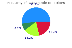
Discount rabeprazole 10 mg without a prescription. How to eat papaya seeds. Uses of papaya seeds.