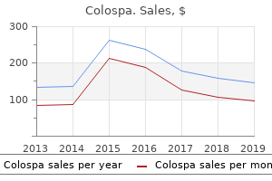
"Order colospa 135 mg without a prescription, muscle relaxant neck pain".
By: O. Rasarus, M.B. B.CH. B.A.O., Ph.D.
Clinical Director, Kansas City University of Medicine and Biosciences College of Osteopathic Medicine
Order genuine colospa on-line
In general spasms near heart colospa 135mg cheap, the degree of pain usually exceeds that anticipated from the clinical findings muscle relaxant potency buy cheap colospa 135mg line. Sinography can be employed to delineate the extent of an abscess cavity or fistula. It is crucial to recognize that necrotizing fasciitis with or without myositis can present as erythema (77%), fluctuance (20%), edema (20%) or induration (43%), clinically resembling a non-fluctuant abscess yet requiring extensive subfascial debridement30. However, severe pain, disproportionate to the physical findings, is present in 94% of cases31. If this complaint is disregarded or misinterpreted as a request for narcotics, the outcome can be devastating. Notably, the classic cutaneous findings of necrotizing fasciitis, such as bullae (noted in 3% of patients), tissue crepitus (3%) and skin necrosis (10%), are only present in a small minority of patients30. For this reason, surgical exploration is mandatory in any parenteral drug user with cellulitis and unexplained severe pain32. Multiple organisms are cultured in 60% to 85% of cases, including anaerobes in 12% of patients. The presence of gas is not pathognomonic for Clostridium infection, as gas gangrene may infrequently be caused by other bacteria. Talc is the main ingredient in some of the narcotic tablets that are crushed, diluted in liquid, and then injected. Talc granulomas may also develop in the lungs, liver, lymph nodes, spleen, and bone marrow. Granulomas may not appear for up to 50 years after subcutaneous injection, because it is the conversion of silica to its colloidal form that stimulates the formation of granulomas34. When the inflammatory nodules ulcerate, they can heal with epidermal pigmentary changes, woody induration of the dermis and subcutaneous tissue, and retracted cicatrices. Mucous Membrane Lesions Snorting cocaine causes erythema of the nasal mucosa, nasal irritation, anosmia and rhinitis, and it may lead to epistaxis, perforation of the nasal septum, and osteolytic sinusitis. A complex consisting of nasal collapse, septal perforation, palatal retraction, and pharyngeal wall ulceration has been described in cocaine snorters. Cuts and blisters on the lips from glass or metal smoking pipes have been noted in addicts who smoke crack cocaine, while ageusia, halitosis and repeated lip smacking are additional signs of cocaine abuse. The labial mucosa may become dry in heroin or amphetamine users12 and amphetamine use may also cause dysgeusia and xerostomia. There are reports of men applying cocaine powder on their glans penis to postpone ejaculation and of women rubbing cocaine on their genitals to enhance pleasure. Penile ulcers have also developed after injections of heroin into the shaft veins. Areca palm seeds (betel nuts) contain the stimulant arecoline and habitual chewing of these nuts, a practice in some areas of Southeast Asia, stains the teeth and is associated with the development of oral leukoplakia and oral submucous fibrosis. The addictive habit of chewing Catha edulis tree leaves (also referred to as khat or qat), which contain the alkaloid stimulant cathinone, is popular in several African and Middle Eastern countries and produces oral keratotic white plaques in ~20% of users. The skin lesions may be discrete papules or erosions that are often confused with acne. Disseminated candidiasis associated with injections of brown heroin was reported in the 1980s and found to be caused by an overgrowth of yeast in the lemon juice used to dissolve the heroin33. Initially, patients developed high fevers, rigors, headaches, myalgias and occasionally jaundice, but, at this stage, blood and urine cultures rarely demonstrated organisms. Most patients eventually developed pustular folliculitis and painful nodules in hairbearing areas (scalp, moustache and beard)33. The lesions resembled bacterial folliculitis but potassium hydroxide examination and lesional biopsy specimens demonstrated the presence of yeast33. The characteristic cutaneous lesion is a cellulitic plaque or abscess with extensive necrotic tissue. Cigarette burns typically occur on the digits and sternum as the cigarette was being held or smoked prior to loss of consciousness12.
Syndromes
- Brain tumor
- Blue lips, nail beds, and skin from lack of oxygen in the blood (cyanosis)
- Antibiotics to treat any infection
- Bleeding from the umbilical cord just after birth
- Other genital or urinary problems
- In some cases, no cause can be found.
- Lung abscess
- Damage to cornea of the eye
Order colospa 135 mg without a prescription
However zanaflex muscle relaxant generic colospa 135mg visa, there have been concerns regarding the adverse side effects of this group of drugs muscle relaxant reversal agents order 135mg colospa mastercard, including hepatotoxicity, weight gain, congestive heart failure, and bone fractures in women. Omega-3 polyunsaturated fatty acids can also lower circulating triglyceride levels by reducing hepatic triglyceride synthesis via competitive inhibition. To date, controlled trials supporting the efficacy of these various drugs in inherited lipodystrophies are lacking. Therapy has also been targeted at the adipokine abnormalities, such as leptin deficiency. Treatment with recombinant methionyl human leptin (r-metHuLeptin) for generalized lipodystrophy, both congenital and acquired forms, has resulted in an improvement of glycemic control, hyperlipidemia, hepatomegaly, and diabetic complications such as proteinuria. The therapy is generally well tolerated, with no notable serious adverse effects60,61. In addition to correcting the metabolic derangements, leptin replacement may improve appetite regulation. However, administration of metHuLeptin has not led to improvement in the lipodystrophy. Because there is activation of the alternative complement pathway, eculizumab has been tried, with mixed results. Insulin Lipodystrophy While the incidence of insulin lipoatrophy has decreased significantly with the advent of purified human insulin, some patients may be injecting other forms. Switching to human insulin and changing the mode of delivery are helpful interventions (see above). For prevention, constant rotation of injection sites, such that the same site is not used more often than once a month, is recommended. Also, with better elucidation of the genetic basis of the various inherited lipodystrophy syndromes (see Table 101. Treatment options for the physical aspects of lipodystrophy are limited (see below). In particular, stavudine and zidovudine were more strongly associated with lipoatrophy, as was didanosine67,68. Interference with the function of the respiratory chain complexes leads to impaired fatty acid oxidation and intracellular accumulation of triglycerides and lactate, which can then enter the systemic circulation. Clinical Features As in hereditary lipodystrophy syndromes, fat redistribution may precede the development of metabolic complications. However, patient self-report, in concert with physician examination, still remains the earliest and best indicator of body shape change. The loss of subcutaneous facial fat, in particular the buccal, parotid and temporal fat, results in prominent zygomata, sunken eyes, deepened and redundant melolabial folds, and a cachectic facies. Central lipohypertrophy presents as: (1) an accumulation of intra-abdominal fat (omental, mesenteric, retroperitoneal), resulting in abdominal protrusion ("protease paunch"; "Crix belly"); and/or (2) increased fat deposition within the dorsocervical fat pad ("buffalo hump"), breasts (gynecomastia in men, larger breasts in women), anterior neck and/or lateral mandibular region, and the muscles and liver. Lipoma formation has also been noted, and the presence of a "buffalo hump" has been shown to be associated with insulin resistance and diabetes mellitus. Although a pilot study had shown that uridine supplementation increased fat mass in patients who continued taking thymidine analogues81, in subsequent randomized studies, no significant improvement in limb fat was observed with uridine supplementation82,83. Facial lipoatrophy can be particularly stigmatizing and injectable fillers can help restore the facial fullness that has been lost. Even though the lipoatrophic process is ongoing, facial volume can be restored by increasing the thickness of the dermis and the effects may last up to 2 years. These fillers are generally safe and well tolerated, with nodule formation being a possible long-term complication. The transfer of autologous fat to the face also leads to improvement, but a donor site may be lacking in patients with significantly reduced body fat. Diet and exercise modifications reduce overall and truncal fat, but as in the general population, adherence to these lifestyle changes is difficult. Use of thiazolidinediones, metformin, and testosterone has not shown any beneficial effect on visceral adipose tissue. Patients with central lipohypertrophy have reduced growth hormone secretion, reduced response to growth hormone-releasing hormone, and increased somatostatin tone (which inhibits growth hormone secretion). As a result of these findings and the known fat-oxidizing and lipolytic properties of growth hormone, the use of daily subcutaneous injections of recombinant growth hormone was investigated. There was some evidence of a reduction in both visceral adipose tissue and abdominal subcutaneous fat; however, its use was associated with a loss of peripheral limb fat and insulin resistance84.
Order cheap colospa online
To avoid confusion muscle relaxant you mean whiskey order 135 mg colospa mastercard, it is recommended that patients be categorized as having multifocal infantile hemangiomas with or without extracutaneous hemangiomas spasms top of stomach cheap colospa 135 mg visa. Multifocal and especially diffuse hepatic hemangiomas are associated with a higher risk of high-output cardiac failure due to arteriovenous or arterioportal shunts, as well as hypothyroidism (see below) and abdominal compartment syndrome related to massive hepatomegaly83. Although hepatic hemangiomas in association with segmental cutaneous hemangiomas have been reported84, no hepatic hemangiomas were found via ultrasound in a prospective study of 60 infants with at least one large (>30 cm2) hemangioma and fewer than five lesions in total85. Segmental cutaneous hemangiomas are occasionally associated with segmental hemangiomas in the gastrointestinal tract, most often in the distribution of the superior mesenteric artery and potentially complicated by bleeding86. Hemangiomas may also develop in a variety of other locations, including mucosal surfaces and the eyes. Increased levels of type 3 iodothyronine deiodinase, an enzyme that deactivates thyroid hormone, have been identified in tissue from proliferating hemangiomas. This can lead to hypothyroidism in infants with large-volume proliferative-phase lesions88. The consumptive nature of the hypothyroidism often makes it difficult to correct, but it resolves as the tumor regresses. Screening for hypothyroidism in the immediate neonatal period is inadequate, as hemangiomas are typically only beginning to develop at that time. Although consumptive hypothyroidism is usually associated with hepatic hemangiomas, it has been described in patients with large hemangiomas in the parotid area, and type 3 iodothyronine deiodinase activity has been noted in cutaneous hemangiomas89. Consequently, it has been recommended that evaluation for hypothyroidism be considered in infants with large cutaneous hemangiomas or hepatic hemangiomatosis88,90. Infants with multiple cutaneous hemangiomas should be monitored for signs or symptoms of visceral involvement with periodic physical examinations. Radiologic evaluation, most often abdominal ultrasonography to screen for hepatic lesions, may be indicated to assess for systemic involvement in some patients. Not all infants with hepatic hemangiomatosis require treatment, but it may be difficult to determine those who will at initial presentation85. It is important to consider the phase of growth when assessing hemangiomas radiographically, because proliferating hemangiomas demonstrate different characteristics than their involuting and involuted counterparts (Table 103. Biopsy specimens obtained from superficial and deep lesions show the same features. Well-defined non-encapsulated masses composed of proliferating plump endothelial cells and pericytes characterize proliferating hemangiomas. Small vascular lumens may be noted focally throughout the tumor, but lumen formation may be more difficult to appreciate in early proliferating lesions. Larger feeding and draining vessels are noted within the septae, and mitotic figures and apoptotic bodies may be present within the tumor mass. An increased number of mast cells is often present within proliferating hemangiomas, and some studies have found even higher numbers of mast cells during early involution40. The involution phase is marked by flattening of the endothelium and reduced numbers of mitotic figures. Eventually, the vessels decrease in number and lose their tightly packed appearance, with fibrous and fatty tissue separating the vessels within and between lobules. These placental markers are also absent in normal vessels of the skin and subcutis19,40. Hemangioma precursors and early proliferating lesions may sometimes be misdiagnosed as capillary malformations or telangiectasias. Other vascular lesions of infancy that may be in the differential diagnosis of superficial hemangioma include tufted angioma, multifocal lymphangioendotheliomatosis with thrombocytopenia, infantile hemangiopericytoma, spindle cell hemangioma, verrucous venous malformation ("verrucous hemangioma"), and eccrine angiomatous hamartoma. Deep hemangiomas provide a greater diagnostic challenge, and radiographic evaluation may be helpful. In addition, some patients with this malignancy present with disseminated intravascular coagulation, which further confuses the clinical picture. However, if the diagnosis is not clarified by clinical and radiographic examinations, or if the lesions are atypical, then histologic examination is indicated. The major goals of management include: (1) preventing or reversing life- or function-threatening complications; (2) treating ulcerations; (3) preventing permanent disfigurement; (4) minimizing psychosocial distress to patients and their families; and (5) avoiding overly aggressive, potentially scarring procedures for lesions that have a strong probability of involuting without significant residua94.
Purchase colospa discount
This produces a characteristic pattern of alternating ortho- and parakeratosis muscle relaxant herbal supplement cheap colospa 135 mg with amex, often referred to as the "flag" or "pink and blue" sign spasms 1st trimester buy genuine colospa line, since the color of the stratum corneum shows a more eosinophilic column (parakeratosis) alternating with a basophilic one (orthokeratosis). Actinic cheilitis arises on the vermilion lips, primarily lower one, and may or may not have associated inflammation. Intercellular bridges (desmosomes) are often apparent, along with keratin pearls and apoptotic cells. The pink quality of the cytoplasm arises from abundant high-molecular-weight keratin. The inflammatory infiltrate varies considerably in intensity and consists primarily of lymphocytes and plasma cells. Only tumors >2 mm in vertical thickness had a significant risk of developing metastases. Other risk factors for metastasis included location on the ear, increased horizontal size, and immunosuppression (Table 108. An inflammatory infiltrate of lymphocytes, and often eosinophils, is usually present. Small intratumoral abscesses of neutrophils are common, and neurotropism may be observed. As the lesion regresses, the dome-shaped architecture flattens and fibrosis develops at the base of the lesion. In the view of the authors, there are four major distinctive clinicopathologic types, namely, nodular, superficial, morpheaform, and fibroepithelial (also referred to as fibroepithelioma of Pinkus)68. Cytologic atypia is minimal, and the tumor has a pushing border as opposed to an infiltrating invasive edge. The major distinguishing features of a verrucous carcinoma are the massiveness and depth of the lesion, as well as the more pronounced irregular architecture. With time, the tumor can enlarge and ulcerate (rodent ulcer, phagedenic ulcer), but an elevated rolled border usually remains and is a clinical clue to the diagnosis. The clinical differential diagnosis of non-ulcerated lesions includes adnexal neoplasms (see Ch. Additional findings include focal scale and/or crusts, a thin rolled border, and variable amounts of melanin; in larger lesions, areas of spontaneous regression may be present, characterized by atrophy and hypopigmentation. Subclinical lateral spread accounts for the significant recurrence rate of these tumors after routine surgical treatment. The clinical differential diagnosis includes solitary lichenoid keratosis and Bowen disease (more scale) as well as inflammatory diseases such as psoriasis, dermatitis, and cutaneous lupus erythematosus. The surface of the lesion is typically smooth, although crusts with underlying erosions or ulcerations as well as superimposed papules may be observed. While an elevated pearly border is typically absent, telangiectasias may be present. The biologic behavior is usually more aggressive, with extensive local destruction. The clinical differential diagnosis includes an intradermal melanocytic nevus or large fibroepithelial polyp (skin tag). It has been estimated that this variant constitutes 1% of all keratinocyte carcinomas. The tumor cells have large, relatively uniform nuclei and scant cytoplasm; cellular borders are indistinct and desmosomes are inapparent. The fibromyxoid stroma is intimately associated with the tumor islands, often showing increased cellularity. In larger tumor islands, central areas of necrosis may develop, leading to the formation of cystic spaces. There is evidence that many of these multifocal buds connect in a net-like pattern, such that most tumors are not truly multifocal.
Safe 135mg colospa
Such products may slow the movements of adult lice and allow them to be more easily combed out of the scalp muscle relaxant yellow pill v buy cheap colospa 135mg online, but these substances are not lethal to lice spasms ms discount colospa online master card. Several essential oils have been reported to be effective as lice therapy, and they have been incorporated into various products that are typically found in health food stores. However, appropriate clinical studies are needed to confirm their safety and efficacy. Many school authorities enforce a "no-nit" policy and do not allow children to return to school if they have nits, regardless of whether they are viable or not. As a result, the tedious task of physically removing all nits via combs with closely spaced metal teeth is required. In comparison to malathion, carbaryl is potentially more toxic to patients, while being less lethal to lice. Topical ivermectin has been shown to kill permethrin-resistant head lice25, and the viability of lice hatched from treated eggs is severely compromised26. In two randomized controlled studies (total n = 765), 74% of patients treated with a single 10-minute application of 0. Oral ivermectin represents another therapeutic option for resistant head lice infestations. In a large multicenter clinical trial, 95% of patients with head lice that previously failed topical therapy (pyrethrin or malathion) who received 400 mcg/kg of ivermectin on days 1 and 8 were lice-free on day 15, compared to 85% of those treated with two applications of topical malathion28. As discussed above for scabies, oral ivermectin is not recommended for children who weigh <33 pounds (15 kg) and pregnant or breastfeeding women. Benzyl alcohol is thought to act via asphyxiation by preventing lice from closing their respiratory spiracles, which become blocked by the lotion; it is not ovicidal. Additional agents that are being developed combine dimethicone with penetrating excipients that may improve its effectiveness. It is a fermentation product of the bacterium Saccharopolyspora spinosa that induces muscle spasms and paralysis in lice when applied topically. Dimethicone Dimethicone is a silicone oil that is used as an emollient in skin care products. Crab lice have serrated edges on their first claw that enable them to ambulate on the entire body surface. Thus, infestation occurs not only in pubic hair, but also in hair of the scalp, eyebrows, eyelashes, moustache, beard, axillae, and perianal area. Indeed, 60% of patients with pubic lice are infested in at least two hair-bearing sites. When the pubic area is shaved or treated, surviving crab lice can travel to other hairy areas of the body, including the scalp. Other findings may include nits at the base of hair shafts, erythema around hair follicles, excoriations, evidence of a secondary bacterial infection, and lymphadenopathy. Macula caerulea are asymptomatic, slate-gray to bluish, irregularly shaped macules that measure 0. These lesions, which typically develop in chronic crab lice infestations, are thought to result from the breakdown of bilirubin to biliverdin by enzymes in louse saliva. In individuals with crab lice, the possibility of additional sexually transmitted infections should be considered and the original source of the infestation sought in order to reduce the risk of recurrence. Introduction Infestation with Pthirus pubis, the crab louse, causes discomfort, pruritus and embarrassment and may coexist with other sexually transmitted infections. History the parasitic relationship between humans and crab lice dates back to prehistoric times. Pathology Crab lice cause nonspecific inflammatory changes in the epidermis and dermis. Because lice live on the surface of the skin, they are not evident histologically.
Knautia arvensis (Field Scabious). Colospa.
- Cough, sore throat, bruises, skin ulcers, eczema, anal fissures and itching, scabies, and roundworm.
- How does Field Scabious work?
- What is Field Scabious?
- Dosing considerations for Field Scabious.
- Are there safety concerns?
Source: http://www.rxlist.com/script/main/art.asp?articlekey=96453
Colospa 135 mg on-line
Pagetoid melanocytosis and cytologic atypia may be present muscle relaxant walgreens purchase discount colospa on line, as well as fibroplasia spasms groin area discount 135 mg colospa visa, a lymphocytic infiltrate, and melanophages. Irregular and confluent junctional nests are often accompanied by cytologic atypia. Many have a mushroom-like polypoidal morphology, with the rather distinctive junctional elements overlying a prominent dermal nevus component. Notably, there are architectural and cytologic features similar to atypical melanocytic nevi. Raised nevi, potentially confused with seborrheic keratoses, are less likely to have a verrucoid surface and pseudo-horn cysts and do not have a cerebriform surface when examined by dermoscopy. Dermatofibromas are usually differentiated from nevi by their very firm consistency, "dimpling" with lateral compression, preference for the lower extremities, and central white patch by dermoscopy. Both neurofibromas and fibroepithelial polyps may be indistinguishable from skin-colored or slightly pigmented, pedunculated intradermal nevi. In general, typical melanocytic nevi are distinguishable from atypical nevi and melanoma by smaller size, overall symmetry and orderly appearance, homogeneous coloration, and regular, well-defined borders. Furthermore, red, blue, gray and black colors are not usually seen in common acquired nevi and should alert one to a potentially atypical lesion. They may display a uniform brown or dark brown color, but often have linear striations. The characteristic dermoscopic features of benign melanocytic nevi of the palms and soles are due to the unique anatomy of acral skin. In benign melanocytic nevi, the nests of nevus cells are situated around the furrows. However, similar nevi may occur in other locations such as the scrotum, perineum, umbilicus, breast, and axilla38. In general, premenopausal women (ages 14 to 40 years) present with this type of vulvar nevus36. These special site nevi appear to be uncommon, perhaps accounting for <10% of nevi removed, but data could be biased by selection factors. In a series of 36 genital nevi from the vulva, perineum and mons pubis, more than half were macular clinically, with the remainder elevated. On occasion, the junctional nests are somewhat enlarged, but they usually have a fairly uniform round or oval shape and are similar in size and spacing. Lentiginous melanocytic proliferation and some degree of upward migration of melanocytes are features commonly observed in acral nevi40. One should be cautious in over-interpreting the latter changes unless prominent architectural disorder and cytologic atypia are also present. The dermal component of acral nevi is often characterized by round nests of melanocytes. The parallelfurrowpatternis evidentwithasmall focusofthelattice pattern Theostiaofthe eccrineductswithinthe epidermalridgescan alsobeseen,especially atthesiteofblueink Epidemiology the prevalence of Spitz nevus in the general population has not been accurately documented41. However, amongst melanocytic lesions that have been surgically excised, approximately 1% exhibit the histologic characteristics of Spitz nevus. Spitz nevi occur in all age groups, but are uncommon beyond 40 to 50 years of age. In one series, 36% of the individuals with Spitz nevi were under the age of 10 years, 33% were between the ages of 10 and 20 years, and 31% were older than 20 years of age41. The association of eruptions of Spitz nevi with pregnancy and puberty suggests that dormant nevi may become hormonally activated. P4) Densely packed, fine parallel lines of pigmentation, often set in a direction that crosses the skin markings Table 112. Relatively flat, polypoid, and pedunculated morphologies have also been described.
Cheap colospa generic
Due to the presence of saprophytic spirochetes in the oral cavity muscle relaxant otc meds purchase cheap colospa, there is limited utility for darkfield examination of serous exudate from lesions in this site bladder spasms 5 year old best colospa 135mg. Immunologic detection of the microorganism by the indirect fluorescent antibody test, which utilizes fluorescein-labeled anti-T. The latter is a component of mammalian cells that is incorporated and modified by treponemes, which results in the development of host antibodies against it (comparable with autoantibodies directed against phospholipids; see Ch. Qualitative non-treponemal tests are suitable for screening purposes, and reactive results have to be confirmed by antibody titer. The performance of a quantitative non-treponemal test is generally requested even with a positive darkfield examination, in order to provide a baseline for longitudinal evaluation after antibiotic therapy. A fourfold decrease in the antibody titer indicates successful treatment, while a fourfold increase indicates relapse or reinfection. In the case of early and efficacious treatment, non-treponemal assays usually become negative. IgM and IgG antibodies are usually detected in these assays by the end of the fourth week after infection. The specific treponemal tests generally remain positive indefinitely, except in the case of treatment of very early syphilis where they do revert to negative. The sensitivity varies with the stage of syphilis: between 70% and 100% in primary syphilis, 100% in secondary and latent syphilis, and about 95% in late syphilis16. A positive result means that the patient had or still has active syphilis, but no assessment about the activity of the disease can be concluded from the qualitative result. Ninety percent of patients are positive at the time they seek medical care for a chancre. To avoid nonspecific reactions with antibodies directed against saprophytic treponemes, a further step with Reiter treponemes is included to absorb nonspecific antibodies. The differentiation into IgM and IgG is possible by including selective anti-Ig antibodies. These tests are useful for the diagnosis of congenital syphilis, neurosyphilis (positive, but low-titer), and reinfection. DifferentialDiagnosis the differential diagnoses for the different stages of syphilis are listed in Table 82. Treatment Penicillin G is still the treatment of choice for all stages of syphilis, and recommended regimens are listed in Table 82. Tetracyclines are used as second-line therapy if penicillin cannot be given; although a single 2 g dose of azithromycin can be effective19, treatment failures due to macrolide-resistant T. Recommended treatment regimens in the settings of pregnancy and congenital syphilis are summarized in Table 82. Evaluation of late syphilis at 6-month intervals for up to 3 years is recommended. Pathology In primary syphilis, there is ulceration and a diffuse dermal infiltrate of plasma cells, lymphocytes, and histiocytes. In secondary syphilis, there is great variability in the histopathological pattern, reflecting the variable clinical appearance of the disease. Older lesions of secondary syphilis may be granulomatous and can resemble sarcoidosis or other granulomatous dermatoses, except for the presence of plasma cells. Endothelial swelling and vascular proliferation can also be seen in secondary syphilis. In tertiary syphilis, tuberculoid granulomas (with or without caseation) are present together with plasma cells. Neonate with a normal physical examination and serum non-treponemal antibody titer less than fourfold the maternal titer; treatment is optional if the mother was fully treated before pregnancy. Neisser", recognizing Albert Neisser, who discovered the microorganism in 1879 in stained smears from vaginal, urethral, and conjunctival exudates.

Cheap colospa online master card
Dosage recommendations back spasms 37 weeks pregnant cheap colospa 135mg, duration of therapy muscle relaxant medicines generic colospa 135 mg, tapering schedules, and monitoring guidelines vary widely. Treatment is usually maintained at these doses until cessation of growth or shrinkage occurs, followed by a gradual taper. A variety of factors influence the taper schedule, including the age of the patient, hemangioma growth rate, reason for treatment, presence of adverse effects, and rebound growth. Doses >3 mg/kg/day resulted in a response rate of 94% but a greater incidence of adverse effects. Concerns have been raised about neurotoxicity in very premature infants treated with corticosteroids for other indications, but there is currently no evidence of similar effects in term infants. Other systemic therapies Vincristine is a chemotherapeutic agent that has been widely used for the treatment of childhood neoplasms. It is a vinca alkaloid that interferes with microtubule formation during mitosis, inducing apoptosis of tumor and endothelial cells. Toxicities include peripheral neuropathy, constipation, jaw pain, and (uncommonly) anemia and leukopenia. Placement of a central venous catheter is required to administer vincristine, and participation of a pediatric oncologist/hematologist is advisable. In vitro treatment of hemangioma endothelial cells with rapamycin results in reduced proliferation18,23. Potential adverse effects include mucositis, hyperlipidemia, headaches, hepatotoxicity, and neutropenia. Multiple treatment sessions, usually every few weeks, are required during the proliferating phase to improve the lesion and prevent rebound growth. Treatments are generally well tolerated, but adverse reactions can include pigmentary alteration, ulceration, and atrophic scarring, although these occur less frequently with the improved cooling technology present in newer generations of pulsed dye lasers147,148. Surgical excision is usually employed for involuted or partially involuted lesions to remove fibrofatty tissue and redundant skin. The optimal timing of surgery depends upon multiple factors, including the location, size, and morphology. Surgical excision during the proliferative phase is controversial and usually reserved for situations in which: (1) a function-threatening hemangioma. A study comparing surgical excision of lip hemangiomas during the proliferative phase versus involution found comparable long-term cosmetic outcomes and less speech delay in those undergoing earlier excision151. Other settings where early surgery may be appropriate include pedunculated lesions for which resection will be inevitable and small, persistently ulcerated lesions that have failed conservative therapy. The ultimate management goal is to achieve as normal an appearance as possible, considering the expected surgical scar as well as the predicted time course and outcome of involution. Arterial embolization has been used to treat life-threatening hemangiomas that cause high-output congestive heart failure, and it may also be utilized in conjunction with other modalities and prior to surgical resection. Small, firm, violaceous papules may be present on the surface, and superimposed milia or hypertrichosis are occasionally noted. Possible complications include necrosis, ulceration, transient thrombocytopenia, and potentially life-threatening hemorrhage that may be heralded by focal crusting3,152,155,156. These lesions may have a slight female predominance and most often develop on the trunk and extremities157. The diagnosis is often established retrospectively when involution fails to occur. Although congenital hemangiomas may be recognized by prenatal ultrasonography during the second and third trimesters, misdiagnosis as other vascular anomalies such as lymphatic or arteriovenous malformations is common160. These include striking lobularity with densely fibrotic stroma, stromal hemosiderin deposits, focal thrombosis and sclerosis of capillary lobules, fewer mast cells, and the coexistence of proliferating vasculature with multiple thin-walled vessels. Most congenital hemangiomas do not require treatment, and medical therapies such as propranolol are generally not effective. Inthis child,multiplesmall, superficialskinlesions werepresentinamiliary patternand hemangiomaswere presentintheliver. Co-segregation of infantile hemangiomas and vascular malformations within families has also been observed.
Buy colospa online from canada
Disfigurement and interference with function because of location Even small lesions may cause complications if they arise in vulnerable locations spasms colon buy 135 mg colospa mastercard. Most commonly muscle relaxant withdrawal symptoms purchase colospa 135mg online, periocular hemangiomas cause astigmatism by compressing the globe and deforming the cornea, which results in asymmetric refractive errors. They may also cause visual abnormalities by obstructing the visual axis or by invading the orbital musculature, which can lead to light-deprivation amblyopia and strabismus, respectively. Lesions located on the upper lid are most problematic, but visual obstruction can complicate lesions on the lower lid as well. Infants with periocular hemangiomas should be evaluated by an ophthalmologist at baseline and closely thereafter during the proliferative stage63,64. Deep and mixed hemangiomas can distort the underlying cartilage and leave significant fibrofatty residua. In rare cases, superficial hemangiomas located along the columella may ulcerate and lead to destruction of underlying cartilage, which may be heralded by the appearance of a horizontal crease in the inferior columella. Lip hemangiomas are often superficial or mixed lesions; painful ulceration is common as they proliferate, which leads to feeding difficulties. Local factors, including recurrent trauma and the commensal bacterial flora, may increase the incidence of this complication. Hemangiomas located on the pinna may ulcerate and become infected, increasing the risk of scarring and distortion of normal structures. Conductive hearing loss can result from obstruction of the external auditory canal by a hemangioma7. These lesions may affect the underlying breast bud, and residual masses may lead to the appearance of breast asymmetry. Early surgical intervention is not advised, as it may ultimately affect normal breast development. Anogenital hemangiomas are often complicated by painful ulceration, and wound care may be difficult in this region. Large hemangiomas of the limbs may also ulcerate, and residual excess tissue and rarely limblength discrepancy can remain upon involution. Extracutaneous involvement Large facial hemangiomas, specifically segmental lesions >5 cm in diameter, can pose challenges beyond the complications noted above. They are often associated with extracutaneous anomalies, especially in female infants66. Occasionally, impaired hearing and endocrine abnormalities such as hypopituitarism and hypothyroidism are additional features69,70. The cerebrovascular anomalies typically occur ipsilateral to the hemangioma and most commonly involve the internal carotid artery, highlighting the need for both head and neck imaging73. Cerebrovascular and cardiovascular changes may be progressive, with potential complications including ischemic stroke75, and affected infants should be followed by neurologists and cardiologists as clinically appropriate. However, airway hemangiomas are occasionally associated with segmental hemangiomas that primarily involve the upper face or small hemangiomas in the "beard" area77. The onset of symptoms, which include noisy breathing and biphasic stridor, ranges from a few weeks to several months of age76. Infants with lower facial hemangiomas of concern should be referred for otolaryngologic evaluation. The risk of spinal dysraphism in an infant or child with an isolated midline lumbosacral hemangioma or residuum of an involuted hemangioma >2. Factors that further increase the risk include a larger or ulcerated hemangioma and the presence of additional cutaneous markers such as a deviated gluteal cleft, lipoma, or skin appendage (see Ch. Evaluation for hepatic involvement is recommended when 5 skin lesions are present. Rarely, infants with a large hemangioma plus <5 small hemangiomas have liver involvement81.
Buy colospa 135 mg
The differential diagnosis of ulceroglandular tularemia includes other ulceroglandular entities muscle relaxant kidney stones order colospa 135mg amex. Staphylococcus muscle relaxant pregnancy safe cheap 135mg colospa with mastercard, Streptococcus, Pasteurella multocida), and infectious causes of a sporotrichoid pattern (see Ch. The previously utilized live attenuated tularemia vaccine is no longer available, and there are ongoing efforts to develop a safe and effective vaccine. It is caused by Klebsiella rhinoscleromatis, a short, immotile Gram-negative coccobacillus that is a subspecies of Klebsiella pneumoniae. Endemic foci exist in Central Europe, Egypt, India, Indonesia, Mexico, Central America, and tropical Africa117. Deficiencies of cellular, but not humoral, immunity cause ineffective phagocytosis by macrophages, giving rise to large, vacuolated, non-lipidized histiocytes with intracellular bacteria (Mikulicz cells)118. Clinical features, diagnostic techniques, the differential diagnosis, and treatment recommendations are outlined in Table 74. Rat-Bite Fever Synonyms: Haverhillfever Sodoku Erythemaarthriticum epidemicum Salmonellosis Salmonellosis refers to the spectrum of infections caused by Gramnegative aerobic bacilli in the genus Salmonella. In contrast, non-typhoidal Salmonella is most commonly acquired from inadequately cooked poultry or eggs as well as other contaminated food products or water, resulting in gastroenteritis. Typhoid fever is characterized by fever, headache, malaise, myalgias, cough, sore throat, nausea, vomiting, diarrhea, and constipation. They occur in up to 30% of Rat-bite fever in North America is most often due to infection with Streptobacillus moniliformis, while infection with Spirillum minus is more common in Asia, where it is known as sodoku. Infection results in an acute illness characterized by fever, arthritis, and a rash. While the disorder is usually caused by a bite (rat-bite fever), it can also occur from close contact with rodents or ingestion of contaminated food, water, or raw milk (Haverhill fever). Although the incidence of rat-bite fever is highest in urban areas with poor sanitation where there is a large population of rats, it can also occur from exposure to pet rats or laboratory rats. Erythema, edema, abscess formation, ulceration, and secondary infection may develop at the site of the bite. It favors the buccal and periorbital areas and is often associated with high fevers, an increased leukocyte count with a left shift, and positive blood cultures. Rose spots often occur in crops during the second to fourth weeks of the illness, and Salmonella spp. Other cutaneous manifestations of salmonellosis include erythema multiforme, Sweet syndrome, hemorrhagic bullae, pustular dermatitis, and a generalized erythematous eruption known as erythema typhosum. Southeast Asia, South America), persistence of fevers, and positive cultures aid in distinguishing typhoid fever from influenza and other viral infections. Current treatment options include quinolones for susceptible strains (with resistance most common in South Asia), ceftriaxone, and azithromycin. One of the hallmarks of the disease is a migratory polyarthritis that occurs in 50% of patients and may mimic rheumatoid arthritis. At 2 to 4 days following the onset of fever and arthritis, most patients with rat-bite fever develop an acrally distributed eruption involving the palms and soles. Morbilliform macules and papules, petechiae, vesicles, pustules, and crusts may be seen. Rat-bite fever can be diagnosed by culturing Streptobacillus moniliformis from blood, synovial fluid or abscess aspirates, which requires special media and conditions; Spirillum minus does not grow in culture. The triad of fever, arthritis/ arthralgias, and a rash can be caused by a variety of infectious diseases, including viral infections.
