Generic serophene 100mg visa
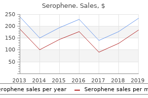
Compartment Models Multicompartment models provide a mathematical framework that can be used to relate drug dose to changes in drug concentrations over time women's health wardenburg generic serophene 100mg with amex. Conceptually senior women's health issues serophene 100 mg on line, the compartments in these models are tissues with a similar distribution time course. The organs and muscles, sometimes called the vessel-rich group, could be the second, or rapidly equilibrating, compartment. Fat and skin have the capacity to bind large quantities of lipophilic drug but are poorly perfused. This is an intuitive definition of compartments, but it is important to recognize that the compartments of a pharmacokinetic model are mathematical abstractions that relate dose to observed concentration. During the distribution phase, the drug moves from the central compartment to the peripheral compartment. In the elimination phase the drug returns from the peripheral compartment to the central compartment and is metabolized and excreted. Without rapid arterial sampling the ultrarapid initial drop in plasma concentration immediately after a bolus injection is missed, and the central compartment volume is blended into the rapidly equilibrating compartment. When rapid arterial sampling is used in pharmacokinetic experiments, the results generally support the use of a three-compartment model. Thus, the number of identifiable compartments reported in a pharmacokinetic study may be more a function of the experimental design than a characteristic of the drug. As previously noted, in compartmental models the instantaneous concentration at the time of a bolus injection is assumed to be the amount of the bolus divided by the central compartment volume. If the bolus is given over a few seconds, the instantaneous concentration is 0, because the drug is all in the vein, still flowing to the heart. More physiologically based models, sometimes called front-end kinetic models, can characterize the initial delay in concentration. The additional complexity that these models introduce is useful only if the concentrations over the first few minutes are clinically important. After the first few minutes, front-end models resemble conventional compartmental models. In the first few minutes following initial bolus administration of a drug the concentration drops very rapidly as the drug quickly diffuses into peripheral compartments. For drugs with very rapid hepatic clearance (eg, propofol) or those that are metabolized in the blood (eg, remifentanil), metabolism contributes significantly to the rapid initial drop in concentration. Following this very rapid drop there is a period of slower decrease in plasma concentration. During this period, the rapidly equilibrating compartment is no longer removing drug from the plasma. The reversed role of the rapidly equilibrating tissues from extracting drug to returning drug accounts for the slower rate of decline in plasma concentration in this intermediate phase. Eventually there is an even slower rate of decrease in plasma concentration, which is log-linear until the drug is completely eliminated from the body. This terminal log-linear phase occurs after the slowly equilibrating compartment shifts from net removal of drug from the plasma to net return of drug to the plasma. Drugs described by two-compartment and three-compartment models will have two or three half-lives. The coefficients A, B, and C represent the contribution of each of the exponents to the overall decrease in concentration over time. The two-compartment model is described by a curve with two exponents and two coefficients, whereas the three-compartment model is described by a curve with three exponents and three coefficients. The mathematical relationships among compartments, clearances, coefficients, and exponents are complex. Every coefficient and every exponent is a function of every volume and every clearance. For drugs described by multicompartment pharmacokinetics (eg, fentanyl, sufentanil), there are multiple elimination half-times, in other words the elimina6 tion half-time is context dependent. Moreover, one cannot easily determine how rapidly a drug effect will disappear simply by looking at coefficients, exponents, and half-lives. For example, the terminal half-life of sufentanil is about 10 h, whereas that of alfentanil is 2 h. This does not mean that recovery from alfentanil will be faster, because clinical recovery from clinical dosing will be influenced by all half-lives, not just the terminal one. Computer models readily demonstrate that recovery from an infusion lasting several hours will be faster when the drug administered is sufentanil than it will be when the infused drug is alfentanil.
Order genuine serophene line
On each side of the external urethral orifice are the openings of the ducts of the para-urethral glands menstrual while pregnant order serophene once a day. The size and appearance of the vaginal orifice vary with the condition of the hymen women's health center lake medina 50mg serophene with mastercard, a thin anular fold of mucus membrane, which partially or wholly 1517 occludes the vaginal orifice. However, its condition (and that of the frenulum of the labia minora) often provides critical evidence in cases of child abuse and rape. They are covered inferiorly and laterally by the bulbospongiosus muscles extending along their length. The greater vestibular glands are round or oval and are partly overlapped posteriorly by the bulbs of the vestibule. The slender ducts of these glands pass deep to the bulbs of the vestibule and open into the vestibule on each side of the vaginal orifice. These glands secrete mucus into the vestibule of the vagina during sexual arousal (see the Clinical Box "Infection of Greater Vestibular Glands"). The lesser vestibular glands are small glands on each side of the vestibule of the vagina that open into it between the urethral and vaginal orifices. These glands secrete mucus into the vestibule, which moistens the labia and vestibule. The internal pudendal artery supplies most of the skin, external genitalia, and perineal muscles. The labial arteries are branches of the internal pudendal artery, as are those of the clitoris. The labial veins are tributaries of the internal pudendal veins and accompanying veins of the internal pudendal artery. Erectile sinus engorgement during the excitement phase of the sexual response causes an increase in the size and consistency of the clitoris and bulbs of the vestibule of the vagina. The anterior aspect of the vulva (mons pubis, anterior labia) is supplied by derivatives of the lumbar plexus: the anterior labial nerves, derived from the ilio-inguinal nerve, and the genital branch of the genitofemoral nerve. Its posterior labial nerves (terminal superficial branches of the perineal nerve) supply the labia. Deep and muscular branches of the perineal nerve supply the orifice of the vagina and superficial perineal muscles. The dorsal nerve of the clitoris supplies deep perineal muscles and sensation to the clitoris (see the Clinical Box "Pudendal and Ilio-Inguinal Nerve Blocks"). In this view, the skin, subcutaneous tissue, and ischio-anal fat bodies have been removed. The bulb of the vestibule and erectile bodies of the clitoris receive parasympathetic fibers via cavernous nerves from the uterovaginal nerve plexus. Parasympathetic stimulation produces increased vaginal secretion, erection of the clitoris, and engorgement of erectile tissue in the bulbs of the vestibule. Lymph from the skin of the perineum, including the anoderm inferior to the pectinate line of the anorectum and the inferiormost vagina, vaginal orifice, and vestibule, drains initially to the superficial inguinal lymph nodes. The operation, usually performed during childhood, removes the prepuce of the clitoris but often also removes part or all of the clitoris and labia minora and may include suturing of the vaginal ostium. This disfiguring procedure is erroneously thought to inhibit sexual arousal and gratification. Vulvar Trauma the mostly vascular bulbs of the vestibule are susceptible to disruption of vessels as the result of trauma. These injuries often result in severe pain, vulvar hematomas (localized collection of blood) in the labia majora, and scarring and, in some cases, may lead to future obstructed labor or fistula formation. Infection of Greater Vestibular Glands 1521 the greater vestibular glands are usually not palpable but are when infected. Occlusion of the vestibular gland duct can predispose the individual to infection of the greater vestibular gland.
Syndromes
- Pieces of the stone block urine flow from your kidney (this may cause severe pain or damage to your kidney)
- Use of humidified air
- Repeated feelings of doubt and failure
- The amount swallowed
- Shiga toxin-producing Escherichia coli (STEC)
- Uterine artery embolization
- Pain in the hip, knee, ankle, and low back
- Low blood pressure (may develop rapidly)
- Side effects of medications
Order serophene 50mg
The dura mater and subarachnoid space (purple) surround the brain and are continuous with that around the spinal cord women's health clinic queenstown serophene 25 mg fast delivery. The two layers of dura separate to form dural venous sinuses menstrual girls buy cheap serophene 100 mg online, such as the superior sagittal sinus. The normal fat- and vein-filled spinal epidural (extradural) space is not continuous with the potential or pathological cranial epidural space. Cranial dura mater has two layers, whereas spinal dura mater consists of a single layer. The calvaria has been removed to reveal the external (periosteal layer) of the dura mater. On the right, an angular flap of dura has been turned anteriorly; the convolutions of the cerebral cortex are visible through the arachnoid mater. The internal aspect of the calvaria reveals pits (dotted lines, granular foveolae) in the frontal and parietal bones, which are produced by enlarged arachnoid granulations or clusters of smaller ones (as in D). The sinuous vascular groove (M) on the lateral wall is formed by the frontal branch of the middle meningeal artery. The intermediate and internal layers (arachnoid and pia) are continuous membranes that collectively make up the leptomeninx (G. This fluidfilled space helps maintain the balance of extracellular fluid in the brain. This fluid leaves the ventricular system and enters the subarachnoid space between the arachnoid and pia mater, where it cushions and nourishes the brain. Dura Mater the cranial dura mater (dura), a thick, dense, bilaminar membrane, is also called the pachymeninx (G. The two layers of the cranial dura are an external periosteal layer, formed by the periosteum covering the internal surface of the calvaria, and an internal meningeal layer, a strong fibrous membrane that is continuous at the foramen magnum with the spinal dura covering the spinal cord. The external periosteal layer of dura adheres to the internal surface of the cranium. Its attachment is tenacious along the suture lines and in the cranial base (Haines, 2013). This outer layer is not continuous with the dura mater of the spinal cord, which consists of only a meningeal layer. The fused external and internal layers of dura over the calvaria can be easily stripped from the cranial bones. The dural infoldings divide the cranial cavity into compartments, forming partial partitions (dural septa) between certain parts of the brain and providing support for other parts. Two sickleshaped dural folds (septae), the falx cerebri and falx cerebelli, are vertically oriented in the median plane; two roof-like folds, the tentorium cerebelli and the small diaphragma sellae, lie horizontally. Venous sinuses of the dura mater and their 1969 communications are demonstrated in the midline vicinity. The tentorium cerebelli is attached along the lengths of 1970 the transverse and superior petrosal sinuses (dashed line). The tentorium cerebelli attaches rostrally to the clinoid processes of the sphenoid, rostrolaterally to the petrous part of the temporal bone, and posterolaterally to the internal surface of the occipital bone and part of the parietal bone. The falx cerebri attaches to the tentorium cerebelli and holds it up, giving it a tent-like appearance (L. The tentorium cerebelli divides the cranial cavity into supratentorial and infratentorial compartments. The supratentorial compartment is divided into right and left halves by the falx cerebri. The brain and part of the calvaria are removed to demonstrate the sinuses related to the falx cerebri and tentorium cerebelli. This view of the interior of the base of the cranium demonstrates most communications of the cavernous sinuses (the inferior communication with the pterygoid venous plexus is a notable exception) and drainage of the confluence of sinuses.

Cheap 50mg serophene amex
As red blood cells move through an artery menopause research order serophene mastercard, a Doppler frequency shift will be detected by the probe menstruation every two weeks cheap 50mg serophene overnight delivery. The difference between transmitted and received frequency causes the characteristic swishing sound, which indicates blood flow. Because air reflects ultrasound, a coupling gel (but not corrosive electrode jelly) is applied between the probe and the skin. Positioning the probe directly above an artery is crucial, since the beam must pass through the vessel wall. Note that only systolic pressures can be reliably determined with the Doppler technique. A variation of Doppler technology uses a piezoelectric crystal to detect lateral arterial wall movement to the intermittent opening and closing of vessels between systolic and diastolic pressure. Auscultation Inflation of a blood pressure cuff to a pressure between systolic and diastolic pressures will partially collapse an underlying artery, producing turbulent flow and the characteristic Korotkoff sounds. These sounds are audible through a stethoscope placed under-or just distal to-the distal third of the blood pressure cuff. Occasionally, Korotkoff sounds cannot be heard through part of the range from systolic to diastolic pressure. This auscultatory gap is most common in hypertensive patients and can lead to an inaccurate diastolic pressure measurement. Korotkoff sounds are often difficult to auscultate in noisy patient care environments and during episodes of hypotension or marked peripheral vasoconstriction. In these situations, the subsonic frequencies associated with the sounds can be detected by a microphone and amplified to indicate systolic and diastolic pressures. When the cuff pressure decreases to systolic pressure, the pulsations are transmitted to the entire cuff, and the oscillations markedly increase. Because some oscillations are present above and below arterial blood pressure, a mercury or aneroid manometer provides an inaccurate and unreliable measurement. A microprocessor derives systolic, mean, and diastolic pressures using an algorithm. Machines that require identical consecutive pulse waves for measurement confirmation may be unreliable during arrhythmias (eg, atrial fibrillation). Nonetheless, the speed, accuracy, and versatility of oscillometric devices have greatly improved, and they have become the preferred noninvasive blood pressure monitors in the United States and worldwide. Arterial Tonometry Arterial tonometry measures beat-to-beat arterial blood pressure by sensing the pressure required to partially flatten a superficial artery that is supported by a bony structure (eg, radial artery). The contact stress between the transducer directly over the artery and the skin reflects intraluminal pressure. Continuous pulse recordings produce a tracing very similar to an invasive arterial blood pressure waveform. Limitations to this technology include sensitivity to movement artifact and the need for frequent calibration. Clinical Considerations Adequate oxygen delivery to vital organs must be maintained during anesthesia. However, flow also depends on vascular resistance: Flow = Pressure Resistance pressure should be viewed as an indicator-but not a measure-of organ perfusion. The narrowest cuff (A) will require more pressure, and the widest cuff (C) less pressure, to occlude the brachial artery for determination of systolic pressure. Whereas the wider cuff may underestimate the systolic pressure, the error with a cuff 20% too wide is not as significant as the error with a cuff 20% too narrow. Incorrect placement or too-frequent cycling of these automated devices has resulted in nerve palsies and extensive extravasation of intravenously administered fluids. In case of equipment failure, an alternative method of blood pressure determination must be immediately available. Contraindications If possible, catheterization should be avoided in smaller end arteries lacking collateral blood flow or in extremities where there is a suspicion of preexisting vascular insufficiency. Invasive Arterial Blood Pressure Monitoring Indications Indications for invasive arterial blood pressure monitoring by catheterization of an artery include A. Selection of Artery for Cannulation Several arteries are available for percutaneous catheterization. Five percent of patients have incomplete palmar arches and lack adequate collateral blood flow. While the operator occludes the radial and ulnar arteries with fingertip pressure, the patient relaxes the blanched hand.
Buy serophene no prescription
Efferent vessels of foot: Venous drainage of the foot primarily follows a superficial route breast cancer in young women order serophene 25mg on line, draining to the dorsum of the foot and then medially via the great saphenous vein or laterally via the small saphenous veins women's health issues who discount 50 mg serophene with visa. The lower limb joints are (A) those of the pelvic girdle connecting the free lower limb to the vertebral column, (B) the knee and tibiofibular joint, and (C) tibiofibular syndesmosis, ankle joint, and the many joints of the foot. During standing, the entire weight of the upper body is transmitted through the hip bones to the heads and necks of the femora. The joint was disarticulated by cutting the ligament of the head of the femur and retracting the head from the acetabulum. The transverse acetabular ligament is retracted superiorly to show the obturator canal, which transmits the obturator nerve and vessels passing from the pelvic cavity to the medial thigh. Except for the depression or fovea for the ligament of the femoral head, all of the femoral head is covered with articular cartilage, which is thickest over weight-bearing areas. The acetabular rim and lunate surface form approximately three quarters of a circle; the missing inferior segment of the circle is the acetabular notch. This superior view of the hip joint demonstrates the medial and reciprocal pull of the peri-articular muscles (medial and lateral 1784 rotators; reddish brown arrows) and intrinsic ligaments of the hip joint (gray arrows) on the femur. Relative strengths are indicated by arrow width: anteriorly, the muscles are less abundant, but the ligaments are robust; posteriorly, the muscles predominate. Parallel fibers linking two discs resemble those making up the tube-like fibrous layer of the hip joint capsule. When one disc (the femur) rotates relative to the other (the acetabulum), the fibers become increasingly oblique and draw the two discs together. Similarly, extension of the hip joint winds (increases the obliquity of) the fibers of the fibrous layer, pulling the head and neck of the femur tightly into the acetabulum, increasing the stability of the joint. In this coronal section of hip joint, the acetabular labrum and transverse acetabular ligament, spanning the acetabular notch (and included in the plane of section here), extend the acetabular rim so that a complete socket is formed. The angle of Wiberg (see text) is used radiographically to determine the degree to which the acetabulum overhangs the head of the femur. Several different lines and curvatures are used in the detection of hip abnormalities (dislocations, fractures, or slipped epiphyses). The Kohler line (red A) is normally tangential to the pelvic inlet and the obturator foramen. A fossa that crosses the line suggests an acetabular fracture with inward displacement. Sectional and radiographic anatomy of gluteal region and proximal anterior thigh at level of hip 1787 joint. Thus, during dissection, the femoral head must be cut from the acetabular rim to enable disarticulation of the joint. This fossa is thin walled (often translucent) and continuous inferiorly with the acetabular notch. In other words, in assuming the upright position, a relatively small degree of joint stability was sacrificed to maximize weight bearing when erect. Even so, the hip joint is our most stable joint, owing also to its complete ball and socket construction (depth of socket), the strength of its joint capsule, and the attachments of muscles crossing the joint, many of which are located at some distance from the center of movement (Palastanga et al. Posteriorly, the fibrous layer crosses the femoral neck proximal to the intertrochanteric crest but is not attached to it. Weight transfer from the vertebral column to the pelvic girdle is a function of the sacro-iliac ligaments. Weight transfer at the hip joint is accomplished primarily by the disposition of the bones, with the ligaments limiting the range of movement and adding stability. Articulating surfaces of hip joint and sites of 1789 attachment and tendinous relationships of iliofemoral ligaments and joint capsule. Because the joint capsule does not attach to the posterior aspect of the femur, the synovial membrane protrudes from the joint capsule, forming the obturator externus bursa to facilitate movement of the tendon of the obturator externus (shown in part C) over the bone. This ligament blends with the medial part of the iliofemoral ligament and tightens during both extension and abduction of the hip joint. The weakest of the three ligaments, it spirals superolaterally to the femoral neck, medial to the base of the greater trochanter. The ligaments and peri-articular muscles (the medial and lateral rotators of the thigh) play a vital role in maintaining the structural integrity of the joint.
Purchase serophene amex
Other pacemaker units must be reprogrammed by placing either an external magnet workout tips women's health buy serophene in india, or womens health 21740 cheap generic serophene canada, preferably, a programming device over the generator. The effect of an external magnet on some pacemakers-particularly during electrocautery-may be unpredictable and should generally be determined prior to surgery. All anesthetic agents have been safely used in patients who already have pacemakers. Local anesthesia with moderate to deep intravenous sedation is usually used for placement of permanent pacemakers. Patient selection for cardiac resynchronization therapy: From the Council of Clinical Cardiology Subcommittee on Electrocardiography and Arrhythmias and the Quality of Care and Outcomes Research Interdisciplinary Working Group in Collaboration with the Heart Rhythm Society. Blood should be immediately available for transfusion if the patient has had previous cardiac surgery (a "redo"); when there has been a previous sternotomy, the right ventricle or coronary grafts may be adherent to the sternum and may be accidentally entered during the repeat sternotomy. Severely compromised patients should be given anesthetic agents in incremental, small doses. Intrathoracic bleeding at a site not adequately drained may cause cardiac tamponade, requiring immediate reopening of the chest. Inadequate surgical control of bleeding sites, incomplete reversal of heparin, thrombocytopenia, platelet dysfunction, hypothermia-induced coagulation defects, undiagnosed preoperative hemostatic defects, or newly acquired factor deficiency or hypofibrinogenemia may be responsible. The period of greatest hemodynamic instability follows the release of the aortic cross-clamp; the abrupt decrease in afterload together with bleeding and the release of vasodilating acid metabolites from the ischemic lower body can precipitate severe systemic hypotension. Surgery on the aorta, the carotid arteries, and the pericardium presents special problems, which are also discussed herein. This technique provides distinctly nonphysiological conditions, because mean arterial pressure is usually less than normal and blood flow is usually nonpulsatile. To minimize organ damage during this stressful period, varying degrees of systemic hypothermia may be employed. Most machines also have separate accessory pumps that can be used for blood salvage (cardiotomy suction), venting (draining) the left ventricle, and administration of cardioplegia solutions. A number of other filters, alarms, and inline pressure, oxygen-saturation, and temperature monitors are also typically used. At the onset of bypass in adults, when using a crystalloid priming solution, hemodilution typically decreases the hematocrit to about 22% to 27%. Blood is included in priming solutions for smaller children and severely anemic adults to prevent excessive hemodilution. A membrane oxygenator permits the perfusion to have independent control of Pao2 and Paco2 by varying the inspired oxygen concentration and the gas flow rate. Heat Exchanger Blood from the oxygenator enters the heat exchanger and can either be cooled or warmed, depending on the temperature of the water flowing through the exchanger; heat transfer occurs by conduction. Because gas solubility decreases as blood temperature rises, a filter or trap is built into the unit to catch any bubbles that may form during rewarming. Thus, the driving force for flow into the pump is directly related to the difference in height between the patient and the reservoir and inversely proportional to the resistance of the cannulas and tubing. Entrainment of air in the venous line can produce an air lock that may prevent blood flow. With some circuits (eg, use of an unusually small venous cannula) assisted venous drainage may be required; a regulated vacuum together with a hard shell venous reservoir or centrifugal pump (see below) is used in 2 such instances. If a "roller" pump is used and the reservoir is allowed to empty, air can enter the main pump and be propelled into the patient where it may cause organ damage or fatality. Centrifugal pumps will not pump air but have the disadvantage of not impelling a well-defined volume with each revolution of the head (unlike roller pumps). Roller Pumps Roller pumps produce flow by compressing largebore tubing in the main pumping chamber as the roller heads turn. The rollers pump blood regardless of the resistance encountered, and produce a nearly continuous nonpulsatile flow. In some pumps, an emergency backup battery provides power in case of an electrical power failure. All roller pumps have a hand crank to allow manual pumping, but those who have hand cranked a roller pump head will confirm that this is not a good long-term solution to an electric power failure. Centrifugal Pumps Centrifugal pumps consist of a series of cones in a plastic housing.
Brittle Willow (Willow Bark). Serophene.
- Treating low back pain.
- Dosing considerations for Willow Bark.
- Osteoarthritis ("wear and tear arthritis"), rheumatoid arthritis, weight loss when taken in combination with other herbs, treating fever, joint pain, and headaches.
- Are there safety concerns?
- Are there any interactions with medications?
- What is Willow Bark?
- How does Willow Bark work?
- What other names is Willow Bark known by?
Source: http://www.rxlist.com/script/main/art.asp?articlekey=96918

100 mg serophene visa
The hypertension associated with laryngoscopy and intubation is often attenuated by intravenous administration of lidocaine (1 breast cancer 14 serophene 25 mg lowest price. Overdoses of lidocaine can lead to marked left ventricular contractile dysfunction breast cancer zumba 50mg serophene otc. Multiple studies have demonstrated that bupivacaine is associated with more pronounced changes in conduction and a greater risk of terminal arrhythmias than comparable doses of lidocaine. Mepivacaine, ropivacaine, and bupivacaine each have a chiral carbon and therefore can exist in either of two optical isomers (enantiomers). The R(+) optical isomer of bupivacaine blocks more avidly and dissociates more slowly from cardiac Na channels than does the S(-) optical isomer (levobupivacaine or ropivacaine). Resuscitation from bupivacaineinduced cardiac toxicity is often difficult and resistant to standard resuscitation drugs. Multiple clinical reports suggest that bolus administration of nutritional lipid emulsions at 1. Onset time and duration of action are similar, but ropivacaine produces less motor block when injected at the same volume and concentration as bupivacaine (which may reflect an overall lower potency as compared with bupivacaine). This improved safety profile likely reflects its formulation as a pure S(-) isomer-that is, having no R(+) isomer-as opposed to racemic bupivacaine. Levobupivacaine, the S(-) isomer of bupivacaine, was reported to have fewer cardiovascular and cerebral side effects than the racemic mixture, but it is no longer available in the United States. Cocaine inhibits the normal reuptake of norepinephrine by adrenergic nerve terminals, thereby potentiating the effects of adrenergic stimulation. Initial treatment of systemic cocaine toxicity should include benzodiazepines to reduce the central stimulation. Cocaine-induced arrhythmias have been successfully treated with -adrenergic antagonists and amiodarone. Cocaine produces vasoconstriction when applied topically and is a useful agent to reduce pain and epistaxis related to nasal intubation in awake patients. Immunological 12 True hypersensitivity reactions (due to IgG or IgE antibodies) to local anesthetics-as distinct from systemic toxicity caused by excessive plasma concentrations-are uncommon. As a consequence, generations of anesthesiologists have speculated whether this preservative may be responsible for most of the apparent allergic responses to amide agents, particularly when skin testing fails to confirm true allergy to the local anesthetic. Compounding the local anesthetic with steroid or epinephrine worsens the myonecrosis. When infused into joints for prolonged periods, local anesthetics can produce severe chondromalacia. Circumoral numbness and apprehension immediately following administration of lidocaine suggest an intravascular injection of local anesthetic. These symptoms and signs after relatively small test doses typically will not be followed by a seizure. She should be closely observed for a possible (but unlikely) seizure and be reassured that the symptoms and signs will soon lapse. The laboring patient is always considered to be at increased risk for aspiration (see Chapter 41); therefore, the airway should be protected by immediate administration of succinylcholine and tracheal intubation (see Case Discussion, Chapter 17). Thus, wherever conduction anesthetics are administered resuscitation drugs and equipment must be available just as for a general anesthetic. Hematological Lidocaine mildly depresses normal blood coagulation (reduced thrombosis and decreased platelet aggregation) and enhances fibrinolysis of whole blood as measured by thromboelastography. These actions could underlie the lower incidence of thromboembolic events in patients receiving epidural anesthetics (in older studies of patients not receiving prophylaxis against deep vein thrombosis). Drug Interactions Local anesthetics potentiate nondepolarizing muscle relaxant blockade in laboratory experiments, but this likely has no clinical importance. As noted earlier, both succinylcholine and ester local anesthetics depend on pseudocholinesterase for metabolism.
Buy serophene 100 mg
Dilation of the annulus of either the mitral or tricuspid valves from ventricular dilation leads to valvular regurgitation menopause 46 100 mg serophene mastercard, further impairing ventricular output menopause xerostomia buy generic serophene online. Increased Sympathetic Tone Sympathetic activation increases release of norepinephrine from nerve endings in the heart and secretion of epinephrine from the adrenal glands into the circulation. Although enhanced sympathetic outflow can initially maintain cardiac output by increasing heart rate and contractility, worsening ventricular function elicits increasing degrees of vasoconstriction in an effort to maintain arterial blood pressure. The associated increase in afterload, however, reduces cardiac output and exacerbates the ventricular failure. Chronic sympathetic activation in patients with heart failure eventually decreases the response of adrenergic receptors to catecholamines (receptor uncoupling), the number of receptors (downregulation), and cardiac catecholamine stores. Nonethe11 less, the failing heart becomes increasingly dependent on circulating catecholamines. Abrupt withdrawal in sympathetic outflow or decreases in circulating catecholamine levels, such as can occur following induction of anesthesia, may lead to acute cardiac decompensation. A reduced density of M2 receptors also decreases parasympathetic influences on the heart. Sympathetic activation tends to redistribute systemic blood flow output away from the skin, gut, kidneys, and skeletal muscle to the heart and brain. Although these mechanisms can initially compensate for mild to moderate cardiac dysfunction, with increasing severity of dysfunction, they may actually worsen the cardiac impairment. Many of the drug treatments of chronic heart failure serve to counteract these mechanisms. Symptoms may also improve in some patients with careful, low-dose -adrenergic blockade. The problem in a pressure-overloaded ventricle is an increase in systolic wall stress. In this case, sarcomeres mainly replicate in parallel, resulting in concentric hypertrophy: the hypertrophy is such that the ratio of myocardial wall thickness to ventricular radius increases. Ventricular hypertrophy, particularly that caused by pressure overload, usually results in progressive diastolic dysfunction. The most common reasons for isolated left ventricular hypertrophy are hypertension and aortic stenosis. He gives a history of having passed out at least once during one of these headaches. Preexcitation usually refers to early depolarization of the ventricles by an abnormal conduction pathway from the atria. The most common form of preexcitation is due to the presence of an accessory pathway (bundle of Kent) that connects one of the atria with one of the ventricles. This abnormal connection between the atria and ventricles allows electrical impulses to bypass the Ventricular Hypertrophy Ventricular hypertrophy can occur with or without dilation, depending on the type of stress imposed on the ventricle. When the heart is subjected to either pressure or volume overload, the initial response is to increase sarcomere length and optimally overlap actin and myosin. With time, ventricular muscle mass begins to increase in response to the abnormal stress. In the volume-overloaded ventricle, the problem is an increase in diastolic wall stress. The increase in ventricular muscle mass is sufficient only to compensate for the increase in diameter: the ratio of the ventricular radius to wall thickness is unchanged. The ability to conduct impulses along the bypass tract can be quite variable and may be only intermittent or rate dependent. Bypass tracts can conduct in both directions, retrograde only (ventricle to atrium), or, rarely, anterograde only (atrium to ventricle). The spread of the anomalous impulse to the rest of the ventricle is delayed because it must be conducted by ordinary ventricular muscle, not by the much faster Purkinje system.
Buy generic serophene 50 mg on-line
The following scenario describes the placement of an internal jugular venous line women's health big book of exercises walmart generic 25 mg serophene free shipping. Central venous catheterization requires full aseptic technique womens health vero beach generic serophene 100 mg overnight delivery, including hand scrub, sterile gloves, gown, mask, hat, bactericidal skin preparation (alcohol-based solutions are preferred), and sterile drapes. A 25-gauge needle is used to infiltrate the apex of the triangle with local anesthetic. Many institutions mandate the use of ultrasound whenever internal jugular vein cannulation is performed. Cannulation of the carotid artery can lead to hematoma, stroke, airway compromise, and possibly death. After free blood flow is achieved we usually confirm central venous versus arterial pressure (using intravenous extension tubing) before introducing a guidewire. We recommend that correct placement of the guidewire be confirmed using ultrasound. The catheter is prepared for insertion by flushing all ports with saline, and all distal ports are "capped" or clamped, except the one through which the wire must pass. The guidewire is removed, with a thumb placed over the catheter hub to prevent aspiration of air until the intravenous catheter tubing is connected to it. Fluid-administration sets should be changed frequently, per your medical center protocol. Blood color and pulsatility can be misleading or inconclusive, and more than one confirmation method should be used. The risks of central venous cannulation include line infection, bloodstream infection, air or thrombus embolism, arrhythmias (indicating that the catheter tip is in the right atrium or ventricle), hematoma, pneumothorax, hemothorax, hydrothorax, chylothorax, cardiac perforation, cardiac tamponade, trauma to nearby nerves and arteries, and thrombosis. Clinical Considerations Normal cardiac function requires adequate ventricular filling. Moreover, right atrial blood oxygen saturation, as opposed to mixed venous saturation (normal is 75%), can be used as an alternative measure to discern tissue oxygen extraction and the adequacy of tissue oxygen delivery. Although echocardiography can readily determine if the heart is full, compressed, contracting, or empty, a trained individual is required to obtain and interpret the images. These measurements might prove particularly important in surgical patients at greatest risk for hemodynamic instability or during surgical procedures associated with a greatly increased incidence of hemodynamic complications (eg, thoracic aortic aneurysm repair). Contraindications 3 Relative contraindications to pulmonary artery catheterization include left bundlebranch block (because of the concern about complete heart block) and conditions associated with a greatly increased risk of arrhythmias. Instead of a central venous catheter, a dilator and sheath are threaded over the guidewire. At approximately 15 cm, the distal tip should enter the right atrium, and a central venous tracing that varies with respiration confirms an intrathoracic position. Transient ectopy from irritation of the right ventricle by the balloon and catheter tip is common and rarely requires treatment. Entry into the pulmonary artery normally occurs by 35 to 45 cm and is heralded by a sudden increase in diastolic pressure. To prevent catheter knotting, the balloon should be deflated and the catheter withdrawn if pressure changes do not occur at the expected distances. Wedging before maximal balloon inflation signals an overwedged position, and the catheter should be slightly withdrawn (with the balloon down, of course). If the latter is suspected, prompt placement of a double-lumen tracheal tube may maintain adequate oxygenation by the unaffected lung. The risk of complications increases with the duration of catheterization, which usually should not exceed 72 h. Optional fiberoptic bundles allow continuous measurement of the oxygen saturation of mixed venous blood. Central line placement should always be completed using rigorous sterile technique, full body draping, and only after multiple, redundant confirmations of the correct localization of the venous circulation.
Cheap 100mg serophene free shipping
The pons is the part of the brainstem between the midbrain rostrally and the medulla oblongata caudally womens healthcare associates generic serophene 25 mg on-line. The medulla oblongata (medulla) is the most caudal subdivision of the brainstem that is continuous with the spinal cord women's health center jackson ms generic 50mg serophene mastercard. It consists of two lateral hemispheres that are united by a narrow middle part, the vermis. Each lateral ventricle opens through an interventricular foramen into the 3rd ventricle. The pyramid-shaped 4th ventricle in the posterior part of the pons and medulla extends inferoposteriorly. It is divided into the posterior cerebellomedullary cistern (cisterna magna) and the lateral cerebellomedullary cistern. Pontocerebellar cistern (pontine cistern): an extensive space ventral to the pons, continuous inferiorly with the spinal subarachnoid space. Interpeduncular cistern (basal cistern): located in the interpeduncular fossa between the cerebral peduncles of the midbrain. Chiasmatic cistern (cistern of optic chiasma): inferior and anterior to the optic chiasm, the point of crossing or decussation of optic nerve fibers. Cisterna ambiens (ambient cistern): located on the lateral aspect of the midbrain and continuous posteriorly with the quadrigeminal cistern (not illustrated). The choroid plexuses consist of fringes of vascular pia mater (tela choroidea) covered by cuboidal epithelial cells. They are invaginated into the roofs of the 3rd and 4th ventricles and on the floors of the bodies and inferior horns of the lateral ventricles. In many places at the base of the brain, only the cranial meninges intervene between the brain and cranial bones. Small, rapidly recurring changes take place in intracranial pressure owing to the beating heart; slow recurring changes result from unknown causes. Momentarily large changes in pressure occur during coughing and straining and during changes in position (erect vs. The bilaterally paired internal carotid and vertebral arteries deliver an abundant supply of oxygen-rich blood. The cervical part of each artery ascends vertically through the neck, without branching, to the cranial base. Each internal carotid artery enters the cranial cavity through the carotid canal in the petrous part of the temporal bone. The orientation drawing (left) indicates the plane of the coronal section that intersects the carotid canal (right). The cervical part of the internal carotid artery ascends vertically in the neck to the entrance of the carotid canal in the petrous temporal bone. The petrous part of the artery turns horizontally and medially in the carotid canal, toward the apex of the petrous temporal bone. It emerges from the canal superior to the foramen lacerum, closed in life by cartilage, and enters the cranial cavity. The artery runs anteriorly across the cartilage; then the cavernous part of the artery runs along the carotid grooves on the lateral side of the body of the sphenoid, traversing the cavernous sinus. Radiopaque dye injected into the carotid arterial system demonstrates unilateral distribution to the brain from the internal carotid artery. A, anterior cerebral artery and its branches; I, the four parts of the 2003 internal carotid artery; M, middle cerebral artery and its branches; O, ophthalmic artery. Armstrong, Associate Professor of Medical Imaging, University of Toronto, Ontario, Canada. The internal carotid and basilar arteries converge, divide, and anastomose to form the cerebral arterial circle (of Willis). The left temporal pole is removed to show the middle cerebral artery in the lateral sulcus of the brain. Clinically, the internal carotid arteries and their branches are often referred to as the anterior circulation of the brain. The anterior cerebral arteries are connected by the anterior communicating artery. The two vertebral arteries are usually unequal in size, the left being larger than the right.
