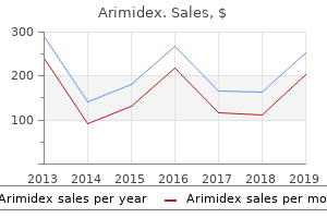
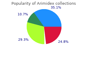
"Buy cheap arimidex on line, womens health johnson city tn".
By: I. Yasmin, M.S., Ph.D.
Clinical Director, Rowan University School of Osteopathic Medicine
Approximately 60% to 70% of the filtered fluid is reabsorbed in the proximal tubule women's reproductive health issues in the philippines discount arimidex online american express. The primary transport process underlying fluid reabsorption is active sodium reabsorption women's health center lansing mi best order for arimidex. Water and many other filtered solutes then are reabsorbed passively as an isosmotic reabsorbate. The Na+ then is extruded actively across the basolateral membrane, giving rise to Na+ reabsorption. Chloride and bicarbonate ion are reabsorbed passively either through the cell or through the tight junctions between the cells (the proximal tubule has "leaky" tight junctions), providing a balance of charge. The tissue is very leaky to water so that as solute is reabsorbed through the cell into the interstitial space, water follows passively by osmotic coupling to the solute, moving through the cells or through the "leaky" tight junctions between the cells. The entry step for Na+ across the luminal cell membrane is normally a coupled process. Both processes are passive steps that are driven by the Na+ gradient into the cell (set up by the Na+ pump). Hence, glucose entry often is referred to as secondary active transport even though the entry step is passive. Once inside the cell, glucose is transported passively across the basolateral membrane by a facilitated diffusion process (passive glucose transporter) into the interstitial space and taken up into the peritubular capillaries. Under normal conditions, all filtered glucose is reabsorbed, except for trace quantities, from the tubular lumen of the proximal tubule, utilizing the Na+glucose cotransport process at the luminal border. However, both Na+ and glucose must bind to specific, but saturable, sites on the Na+-glucose cotransport carrier protein, making glucose reabsorption saturable. Hence, under conditions of elevated plasma glucose levels, such as in diabetes mellitus, or an increased glomerular filtration rate, such as in pregnancy, the filtered glucose load can exceed the capacity for glucose transport; that is, the Na+ glucose cotransporters become saturated, leaving un-reabsorbed glucose behind in the tubular fluid which is swept away into the final urine (glycosuria). In the presence of un-reabsorbed glucose, the "trapped" glucose will act as an osmotic solute, leading to an osmotic diuresis. The associated diuresis can be particularly problematic in patients with diabetes mellitus. The reabsorption of other organic solutes in the proximal tubule also is coupled to Na+ as a Na+-solute cotransporter at the luminal cell membrane, Both sugars, such as galactose, and most amino acids, such as glutamate and glycine, are cotransported with Na+ and display both a tubular transport maximum and a renal plasma threshold (the transport of some amino acids, such as lysine and proline, is not Na+ dependent). Galactose can compete with glucose for binding and transport by the Na+-glucose carrier so that with elevated plasma levels of galactose, such as in pregnancy, galactose can contribute to appearance of glucose in the urine. The proximal tubule is also the site of reabsorption of certain organic acids, with the most dominant normally being lactate anion. Two Na+dependent cotransport process at the luminal membrane appear to underlie organic acid reabsorption: one specific for monocarboxylates such as lactate, pyruvate, acetoacetate, and -hydroxybutyrate and the other for di- and tricarboxylates such as malate, succinate, and citrate. Once inside the cell, the carboxylates exit the cell by means of a variety of exchange processes. Other organic acids, such as urate, an end product of purine catabolism, are both secreted and reabsorbed in the proximal tubule; both processes are Na+-independent, with the reabsorptive mechanisms dominating. Finally, the proximal tubule is the site of secretion of numerous organic anions (paraaminohippurate, oxalate) and cations (choline, guanidine) by separate, saturable transport processes that often involve anion exchange processes that are Na+-independent. Larger peptides and proteins such as myoglobin and albumin bind to the luminal membrane and enter the cell by receptor-mediated endocytic processes and are delivered to lysosomes for degradation. Some filtered inorganic anions, such as sulphate and phosphate, also are reabsorbed in the proximal tubule via a Na+ cotransport process; hence, their transport can be defined by a Tm and renal plasma threshold. The appearance of glucose in the urine is a consequence of which of the following processes in the proximal tubule Upon admission to the emergency room, he is diagnosed with diabetic ketoacidosis, which is accompanied by extreme hypovolemia, supposedly because of the brisk diuresis. His urine is "tea" colored as a result of breakdown of skeletal muscle by the cocaine, so-called rhabdomyolysis. In diabetes mellitus, in which plasma glucose levels are markedly elevated, the high glucose load being filtered (with an elevated concentration) can exceed the capacity of the luminal Na+-glucose cotransporter to reabsorb glucose (ie, the carrier is saturated). The excess glucose that is not reabsorbed is trapped in the tubular fluid because no transport pathways are present in later nephron segments to reabsorb this hexose.
A menopause vs pregnancy symptoms purchase arimidex 1mg without a prescription, Color fundus photograph revealing an area of intraretinal disorders with overlapping clinical features that share in whitening corresponding to a supratemporal common the presence of discrete pregnancy 9 months generic arimidex 1 mg with visa, multiple, wellbranch artery occlusion in the left eye. Common presenting symptoms include photopsias, blurred vision, nyctalopia, floaters, and visual field loss contiguous with the blind spot. Other than patients with birdshot uveitis or serpiginous choroiditis, the majority of individuals are younger than 50 years of age. An increased prevalence of systemic autoimmunity both in patients with white dot syndromes and their first- and second-degree relatives suggests that inflammatory chorioretinopathies may occur in families with inherited immune dysregulation that predisposes to autoimmunity. Whether the white dot syndromes represent a clinical spectrum of a single disease entity or are each discrete diseases awaits identification of the underlying mechanisms. Although they have similarities, the white dot syndromes can be differentiated by their variable lesion morphology and evolution, distinct natural histories, and appearance with multimodal imaging. This differentiation has important implications with respect to disease-specific treatments and visual prognosis. White dot syndromes: a 20-year study of incidence, clinical features, and outcomes. White spot syndromes of the retina: a hypothesis based on the common genetic hypothesis of autoimmune/inflammatory disease. Increased prevalence of autoimmunity in patients with white spot syndromes and their family members. Birdshot uveitis Birdshot uveitis (also known as birdshot retinochoroidopathy, birdshot chorioretinopathy, and vitiliginous chorioretinitis) is an uncommon disease presenting predominantly in white women of northern European descent past the fourth decade of life. The presence of the haplotype confers considerable increased relative risk (224-fold) for the development of this disease. Presenting symptoms include blurred vision, floaters, nyctalopia, and disturbance of color vision. Visual complaints can be out of proportion to the measured Snellen visual acuity, reflecting the diffuse retinal dysfunction that occurs in this entity. Patients may also report unusual peripheral visual phenomena, such as pinwheels, sparkles, or flickering lights, and these symptoms may be indicators of subtle disease activity. Anterior segment inflammation may be minimal or lacking; however, varying degrees of vitritis are commonly noted. The lesions do not become pigmented over time and are best appreciated by indirect ophthalmoscopy. The characteristic lesions may not be readily apparent at first, and the disorder may be misdiagnosed initially as idiopathic intermediate or posterior uveitis. Fluorescein angiography reveals inconsistent findings depending on age, lesions, and phase of study. Late hypopigmented lesions typically do not show transmission defects, implying loss of pigment concurrent with loss of choriocapillaris. It can also demonstrate patchy or diffuse loss of photoreceptors (inner/outer segment line or ellipsoid zone) and macular thinning, especially with long-standing disease. It is crucial for clinicians to recognize that this entity can be insidious and understand that simply monitoring visual acuity and clinical examination findings is insufficient to protect patients from vision loss. A subset of patients with birdshot uveitis may have self-limited disease and do well without treatment. However, there is no way to determine in advance which patients will have disease progression and which will not, so any patient who is not treated should be monitored closely using the modalities discussed above. Although older studies demonstrated high rates of vision loss in patients who were either not treated or treated in a limited or intermittent fashion, a more recent study indicates that up to 88% of patients can maintain vision with aggressive, long-term control of inflammation. Birdshot uveitis is typically incompletely responsive to corticosteroids alone, and extended treatment is anticipated in most patients given the chronic nature of the disease. The intravitreal fluocinolone acetonide implant is an option for patients who cannot tolerate systemic therapy or in whom systemic therapy has failed. A, Color fundus characterisation and monitoring in the management of birdshot photograph showing multifocal hypopigmented chorioretinopathy.

Baseline variability menstruation quotes funny purchase arimidex australia, which is best assessed with scalp electrodes menstrual pain icd 9 buy arimidex pills in toronto, has become an important sign of fetal well-being and represents a normally functioning autonomic system. Central nervous system depressants (opioids, barbiturates, volatile anesthetics, benzodiazepines, or magnesium sulfate) and parasympatholytics (atropine) also decrease baseline variability, as do prematurity, fetal arrhythmias, and anencephaly. By 32 weeks, fetuses display periodic increases in baseline heart rate that are associated with fetal movements. The mechanism is thought to involve increases in catecholamine secretion with decreases in vagal tone. Accelerations diminish with fetal sleep, some drugs (opioids, magnesium, and atropine), as well as fetal hypoxia. Accelerations to fetal scalp or vibroacoustic stimulation are considered a reassuring sign of fetal well-being. The absence of both baseline variability and accelerations is nonreassuring and may be an important sign of fetal compromise. Three parameters are evaluated: baseline heart rate, baseline variability, and the relationship to uterine contractions (deceleration patterns). Monitoring of heart rate is most accurate when fetal scalp electrodes are used, but this may require rupture of the membranes and is not without complications (eg, amnionitis or fetal injury). An increased baseline heart rate may be due to prematurity, mild fetal hypoxia, chorioamnionitis, maternal fever, maternally administered Deceleration Patterns A. Early decelerations are generally not associated with fetal distress and occur during descent of the head. They are thought to represent decreased arterial oxygen tension on atrial chemoreceptors. Late decelerations with normal variability may be observed following acute insults (maternal hypotension or hypoxemia) and are usually reversible with treatment. Late decelerations with decreased variability are associated with prolonged asphyxia and may be an indication for fetal scalp sampling (see Other Monitoring section below). Complete abolition of variability in this setting is an ominous sign signifying severe decompensation and the need for immediate delivery. Aortocaval compression, maternal hypoxemia or hypotension, or excessive uterine activity (during oxytocin infusions) must be corrected. Changes in maternal position, supplemental oxygen, and intravenous ephedrine or fluid, or adjustments in an oxytocin infusion often correct the problem. Failure to relieve fetal stress, as well as progressive fetal acidosis and asphyxia, necessitate immediate delivery. General Care of the Neonate One healthcare provider whose sole responsibility is to care for the neonate and who is capable of providing resuscitation should attend every delivery. As the head is delivered, the nose, mouth, and pharynx are suctioned with a bulb syringe. After the remainder of the body is delivered, the skin is dried with a sterile towel. Once the umbilical cord stops pulsating or neonatal breathing is initiated, the cord is clamped and the neonate is placed in a radiant warmer with the bed tilted in a slight Trendelenburg position. If the neonate is obviously depressed, the cord is clamped early and resuscitation is initiated immediately. Respirations are assessed by auscultation of the chest, whereas heart rate is determined by palpation of the pulse at the base of the umbilical cord or auscultation of the precordium. In addition to respirations and heart rate, color, tone, and reflex irritability should be evaluated. The 1-min score correlates with survival, whereas the 5-min score has limited relationship to neurological outcome. These decelerations are variable in onset, duration, and magnitude (often >30 beats/ min). They are typically abrupt in onset and are thought to be related to umbilical cord compression and acute intermittent decreases in umbilical blood flow.
Angle closure from mechanisms other than pupillary block also does not require iridotomy (ie pregnancy exercise plan arimidex 1mg without a prescription, neovascular glaucoma and iridocorneal endothelial syndrome) breast cancer 10 year survival rate cheap arimidex 1mg without prescription. An eye with active rubeosis iridis may bleed and develop a large hyphema following laser iridotomy. The risk of bleeding is also increased in a patient taking systemic anticoagulants, including aspirin. Preoperative considerations In acute angle closure, performing laser iridotomy is often difficult due to the cloudy cornea, shallow chamber, and engorged iris that are present in this condition. Corneal edema may be improved prior to laser iridotomy by pretreatment with topical glycerin. In prophylactic iridotomies, pretreatment with pilocarpine may be helpful by stretching and thinning the iris. The patient should be asked about anticoagulants, as their use increases the risk of hyphema. Laser iridotomy or surgical iridectomy breaks the pupillary block and results in opening of the entire peripheral angle (bottom) if no permanent peripheral anterior synechiae are present. The argon laser alone can be used for performing iridotomy in most eyes, but very dark and very light irides present technical challenges. There are variations in technique, and iris color dictates which technique is chosen. Often, compression of the eye with the laser lens will provide a tamponade for the vessel, thereby slowing bleeding until coagulation can occur. In rare cases when this does not work, it may be helpful to use an argon laser to coagulate the vessel. Topical corticosteroids are usually prescribed for 1 week, longer if necessary, as prophylaxis against inflammation. The effects of iridotomy size and position on symptoms following laser peripheral iridotomy. Dysphotopsia after temporal versus superior laser peripheral iridotomy: a prospective randomized paired eye trial. Laser Gonioplasty, or Peripheral Iridoplasty Indications Gonioplasty, or iridoplasty, is a technique to deepen the angle. It is primarily used in persistent appositional angle-closure glaucoma after successful iridotomy in cases of plateau iris syndrome, nanophthalmos, and lens-related angle-closure. It is also used in cases of acute angle closure in which a shallow chamber precludes iridotomy. Stromal burns are made in the peripheral iris with the argon laser to cause contraction and flattening, thereby pulling the iris away from the angle. Contraindications the contraindications for gonioplasty are similar to those for laser iridotomy but also include tumors of the iris or ciliary body and uveitis. Preoperative considerations An angle that is appositionally closed from plateau iris syndrome will not open after laser iridotomy because forward displacement of the ciliary processes pushes the peripheral iris into the drainage angle. Technique Long duration and large spot size with relatively low power is necessary to cause a contraction burn that will thin peripheral iris and pull it out of the angle. Pilocarpine is given preoperatively to maximally stretch the iris, and a contact lens with a gonioscopy mirror is placed on the eye. Postoperative anterior uveitis is common and should be treated with topical corticosteroids. The most common modalities in current practice are endoscopic cyclophotocoagulation and diode laser transscleral cyclophotocoagulation. Endoscopic cyclophotocoagulation is an intraocular procedure in which a microendoscope applies laser energy to the ciliary processes under direct visualization. In transscleral cyclophotocoagulation, the laser probe is placed externally, which focuses the beam across the sclera to cause destruction of the underlying ciliary body and ciliary epithelium. The risk of hypotony and phthisis bulbi is much lower with these latter modalities (unless the eyes are ischemic), and these modalities have been safely used in eyes with good vision. The procedure is useful in all types of glaucoma, and it can be considered for elderly patients when other glaucoma surgeries are refused or not possible because of poor systemic health.
