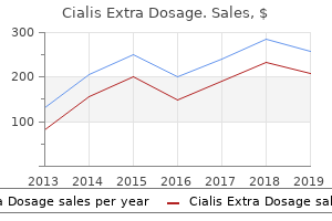
Buy discount cialis extra dosage
The flexibility of the ureteroscope decreases when an instrument is present in the working channel; however erectile dysfunction treatment pune cialis extra dosage 60 mg discount, small-diameter holmium laser fibers have been developed that are both flexible and durable erectile dysfunction in diabetes mellitus pdf purchase cialis extra dosage 50mg with mastercard, causing only minimal resistance during deflection. Catheter inserted over guidewire for contrast injection; guidewire removed Sheath C. Distal sensor flexible ureteroscope inserted in to sheath and advanced in to renal pelvis Before undergoing ureteroscopy, the patient should have a documented negative urinalysis and urine culture, so as to reduce the risk of urosepsis. The majority of ureteroscopic procedures are performed in a specialized cystoscopy suite. The patient is placed in a dorsal lithotomy position, with the lower extremities in stirrups. The procedure is typically initiated by visualizing the bladder lumen with a cystoscope (see Plate 10-37) and then deploying a guide wire in to the ureteric orifice. The guidewire may be placed with either a rigid or flexible cystoscope, depending on surgeon preference. Next, a ureteral catheter is inserted over the wire, and a retrograde pyeloureterogram is performed to evaluate the anatomy of the upper tract and provide a map for deployment of the ureteroscope. The wire, which provides a map of the upper urinary tract, can remain in place throughout the procedure. Thus before deployment of a flexible ureteroscope, a second guide wire is typically inserted to act as a "safety" wire, which remains present throughout the entire procedure and provides access to the upper urinary tract should normal anatomy become disrupted. To place a safety wire, a coaxial dilator/ sheath is introduced over the first wire. The inner dilator is removed, the safety wire is introduced through the sheath, and then the sheath is removed. When a flexible ureteroscope is being used, a ureteral access sheath can be placed early on to facilitate multiple insertions of the ureteroscope and limit the trauma associated with each passage. These sheaths also facilitate drainage of irrigation fluid, therefore permitting more frequent flushing of stone fragments and other debris created during lithotripsy procedures. A tapered inner obturator in its lumen facilitates its passage through the ureter and helps dilate narrowed regions that would otherwise be difficult to traverse. As the ureteroscope is advanced to the desired position, fluoroscopy is performed to monitor its progress in real time. Throughout the process, the urinary tract is irrigated with saline to facilitate ureteroscope passage and improve visualization. Irrigation pressure can be controlled by gravity, a compression bag, or hand-held pumps. If passage of the ureteroscope is difficult, the ureter may be dilated by passing a balloon dilator over the guide wire. Once the ureteroscope has reached the level of interest, various instruments can be introduced in to the working channel to perform a diagnostic. At the end of the procedure, a ureteral stent should be deployed if a ureteral access sheath has been used because the latter is associated with a risk of mucosal injury and postoperative ureteral edema. Ureteral stents are typically biocompatible polyethylene or silicone polymer devices. Most stents have curls at their proximal and distal ends, which help anchor them in the renal pelvis and bladder. In addition, most stents have small holes along their shaft to facilitate drainage. A ureteral stent may be placed through the working channel of a rigid cystoscope, or it can be deployed over a wire using fluoroscopic guidance. A plastic tube known as a stent pusher is used to ensure that the proximal curl reaches the renal pelvis. After fragmentation is complete, the laser is withdrawn, and a stone basket is introduced in to the working channel. The basket is used to remove the larger fragments, which are less likely to be spontaneously voided. The most common complications include stent colic (discomfort from the ureteral stent), transient hematuria, and urinary tract infection. Most cases of ureteral perforation, however, successfully heal with stent deployment alone. Ureteral avulsion is a rare complication of ureteroscopy that is most often repaired using open surgical technique. Ureteroscopy is typically performed on an outpatient basis and does not require hospital admission.
Sweet Bay. Cialis Extra Dosage.
- Are there any interactions with medications?
- Cancer, dandruff, and relieving gas.
- How does Sweet Bay work?
- Dosing considerations for Sweet Bay.
- Are there safety concerns?
- What is Sweet Bay?
Source: http://www.rxlist.com/script/main/art.asp?articlekey=96674
60mg cialis extra dosage for sale
It is then conveyed along axons to the posterior pituitary for storage and release erectile dysfunction epocrates cheap cialis extra dosage 50mg mastercard. These receptors erectile dysfunction support groups trusted 40 mg cialis extra dosage, located outside of the blood-brain barrier, are extremely sensitive to changes in plasma osmolality. Their activation has been hypothesized to occur when there is a loss of intracellular fluid secondary to increased extracellular osmotic pressure. In support of this hypothesis, the osmoreceptors are not equally sensitive to all solutes. Sodium, for example, reliably activates osmoreceptors at high concentrations because, as a predominantly extracellular ion, it establishes a transmembrane osmotic gradient. In contrast, urea and glucose generally do not activate osmoreceptors even at high concentrations because they freely enter cells, thus failing to establish an osmotic gradient. When patients experience extreme insulin depletion, however, osmoreceptors may become sensitive to high concentrations of glucose, presumably because of its increased restriction to the extracellular space. In this setting, the primary objective is to retain intravascular volume, rather than to adjust plasma osmolarity. Such release is mediated by baroreceptors in the atria, aorta, and carotid sinus, which send afferent signals to the brain along the vagus and glossopharyngeal nerves. This sensing mechanism is not nearly as sensitive as the osmolality-sensing apparatus, however, and does not become active until 5% to 10% of plasma volume has been lost. As water is reabsorbed in the cortical and % Change in blood volume or pressure outer medullary collecting duct, urea becomes increasingly concentrated in the tubular lumen. The collecting duct becomes permeable to water, which is reabsorbed because of the high osmotic pressure generated by the solute concentrated in the medullary interstitium. Both the sensor and effector limbs of this feedback circuit reside in the juxtaglomerular apparatus, located where the distal tubule of a nephron contacts its parent glomerulus. As described on Plate 1-20, the juxtaglomerular apparatus contains the macula densa (located in the thick ascending limb), terminal afferent arteriole, initial efferent arteriole, and extraglomerular mesangium. When tubular flow rates are reduced, for example, the macula densa triggers renin secretion from granular cells, which are located in the walls of the terminal afferent arteriole and initial efferent arteriole. Renin release has multiple effects, described later in detail, that promote volume retention and systemic vasoconstriction. In addition to signals from the macula densa, several additional factors can also modulate both afferent arteriolar tone and renin release. A rise in renal perfusion pressure, for example, causes stretching of the afferent arteriole, which increases calcium influx in to smooth muscle and granular cells. Meanwhile, increased sympathetic tone, as occurs during volume depletion, leads to afferent arteriolar vasoconstriction, which redirects blood toward organs with high oxygen extraction (brain, heart, skeletal muscle), as well as activation of renin release. Sympathetic nerves Stimulate afferent arteriolar constriction and release of renin-filled vesicles from granular cells Macula densa Increased Na /Cl reabsorption stimulates afferent arteriolar constriction and suppresses renin release from granular cells. Diminished reabsorption stimulates afferent arteriolar vasodilation and promotes renin release. Extraglomerular mesangial cells (polkissen, Lacis cells) Likely act as signaling intermediaries between the macula densa and granular cells Stimulus Increased tubular flow Effect Afferent arteriolar constriction Suppression of renin release Afferent arteriolar dilation Activation of renin release Afferent arteriolar constriction Suppression of renin release Afferent arteriolar constriction Activation of renin release Decreased tubular flow Afferent arteriole stretching Sympathetic tone the available evidence suggests that the macula densa, located at the end of the thick ascending limb, senses tubular flow based on the concentrations of sodium and chloride in the local filtrate. When tubular flow rates are high, there is a slight decrease in solute reabsorption before the macula densa, and thus higher concentrations of sodium and chloride are present at this area. Adenosine, in turn, activates receptors on the surface of nearby extraglomerular mesangial cells, causing an increase in intracellular calcium levels. A wave of intracellular calcium is transmitted across gap junctions to the smooth muscle and granular cells of the afferent and efferent arterioles, causing constriction of the afferent arteriole and inhibition of renin release. In acute tubular necrosis, for example, there is damage to the proximal tubule, which increases the electrolyte load delivered to the macula densa. As a result, there is severe afferent arteriolar constriction, which is likely a major cause of the decreased filtration function that accompanies this condition. In the early stages of diabetic nephropathy (see Plate 4-46), chronic glucosuria leads to increased glucose-mediated proximal tubular reabsorption, which decreases the electrolyte load delivered to the macula densa. As a result, there is afferent arteriolar vasodilation, leading to hyperfiltration. Because this hormonal network is activated in response to renal hypoperfusion, its effects raise systemic blood pressure through expansion of extracellular volume and systemic vasoconstriction.
Buy cialis extra dosage with mastercard
Although skin metastasis often arises in the vicinity of the underlying primary malignancy erectile dysfunction age 33 purchase cialis extra dosage overnight, the location of tumor metastases is not a reliable means of predicting the primary source erectile dysfunction etiology order cialis extra dosage 100 mg with visa. Sister Mary Joseph nodule is a name given to a periumbilical skin metastasis from an underlying abdominal malignancy. This has been described to occur most commonly with ovarian carcinoma, gastric carcinoma, and colonic carcinoma. Cutaneous metastasis from melanoma can manifest with the rapid onset of multiple black papules and macules that continue to erupt. It is believed to be caused by the systemic production of melanin with deposition in the skin. Breast carcinoma is another form of malignancy that frequently metastasizes to the skin. Breast carcinoma tends to affect the skin within the local region of the breast by direct extension. Pathogenesis: the exact reason why some tumors metastasize to the skin is unknown. Metastases are likely to be dependent on size, ability to invade surrounding tissues (including blood and lymphatic vessels), and ability to grow at distant sites far removed from the original tumor. Histology: the diagnosis of cutaneous metastasis is almost always made by the pathologist after histological review. The risk of recurrence is high, and adjunctive chemotherapy and radiotherapy should be considered. The overall survival rate for multiple cutaneous metastases has been reported to be 3 to 6 months. The tumor is derived from the dermal fibroblast, and it is not believed to arise from previously existing dermatofibromas. Dermatofibrosarcoma protuberans rarely metastasizes, but it has a distinctive tendency to recur locally. Clinical Findings: Dermatofibrosarcoma protuberans is a slow-growing, locally aggressive malignancy of the skin. These tumors are low-grade sarcomas and make up approximately 1% of all soft tissue sarcomas. The tumor is found equally in all races and affects males slightly more often than females. Most tumors grow so slowly that the patient is not aware of their presence for many years. It slowly infiltrates the surrounding tissue, particularly the subcutaneous tissue. If the tumor is allowed to grow long enough, the malignancy will grow in to the fat and then back upward in the skin to develop satellite nodules surrounding the original plaque. This rapid growth phase allows the tumor to grow in a vertical direction, and hence the term protuberans is applied. If medical care is not undertaken, the tumor will to continue to invade the deeper structures, eventually invading underlying tissue, including fascia, muscle, and bone. Dermatofibrosarcoma protuberans is, for the most part, asymptomatic in the initial phases of the tumor. As it enlarges, the patient may notice an itching sensation or, less frequently, a burning sensation or pain. As the tumor enlarges, patients often notice tightness of the skin or a thickening sensation; however, this development is so slow that most patients ignore it for many more months or even years. The differential diagnosis is often between dermatofibrosarcoma protuberans and a keloid or hypertrophic scar. One clue to the diagnosis of dermatofibrosarcoma is the loss of hair follicles within the tumor region. If the tumor is allowed to enlarge enough, it will begin to outgrow its blood supply, and ulceration and erosions develop thereafter.
Cheap cialis extra dosage 200 mg mastercard
It is likely that many patients do not seek medical advice because the onset is insidious or the area of involvement is so small that it is hardly noticeable or bothersome ginkgo biloba erectile dysfunction treatment cheap cialis extra dosage master card. As it expands erectile dysfunction pills australia cheap cialis extra dosage 40mg with mastercard, the central portion becomes slightly hypopigmented and indurated in nature. If the involved area crosses over a joint, there may be some loss of motion of the affected joint and pain with flexion and extension. The main differential diagnosis is between morphea and lichen sclerosis et atrophicus. Lichen sclerosis et atrophicus is typically more strikingly white in coloration and is less indurated. Guttate morphea manifests with tiny, teardrop-shaped areas of hypopigmented macules with slight induration scattered about the trunk or extremities. The induration of guttate morphea is not nearly as prominent as that of localized morphea and may not be appreciable. These guttate lesions may be impossible to distinguish clinically from lichen sclerosis et atrophicus, and a biopsy is the only way to differentiate the two. Generalized morphea is a rare variant with extensive involvement of the cutaneous surface. By definition, generalized morphea does not have systemic involvement, differentiating it from progressive systemic sclerosis. However, patients with generalized morphea may develop atrophy of the adipose and muscle tissues underlying the areas of involvement. Linear morphea, also called linear scleroderma, is a unique cutaneous variant that is well described and has a distinctive appearance and potential underlying complications. The affected skin may become bound down and cause limb length discrepancies as the child grows. Cortical hyperostosis of the long bones underneath the area of linear morphea has been well reported and is termed melorheostosis. There are subtypes of linear morphea that have been given the names en coup de sabre and Parry-Romberg syndrome. En coup de sabre is a specific type of morphea that occurs along the forehead, as well as partially on to the cheek and in to the scalp. It appears as a depressed linear furrow from the scalp vertically down the forehead. The appearance can be subtle or extremely noticeable and can cause significant cosmetic problems. Parry-Romberg syndrome is a name given to linear morphea that occurs vertically across the face, causing hemifacial atrophy. The underlying adipose tissue, muscle, and bone are involved, with significant disfigurement. Often surronded by a violaceous or erythematous rim Progressive systemic sclerosis (scleroderma). Typical skin changes in scleroderma: extensive collagen deposition and some epidermal atrophy reaction in which an excessive amount of collagen is produced locally by fibroblasts. Potential factors that may initiate the reaction are endothelial damage, certain Borrelia burgdorferi infections, and fibroblast abnormalities that lead to increased collagen production. Borrelia-induced morphea has yet to be described in the United States; it has been reported in Europe and Asia. Histology: A punch biopsy specimen of morphea appears as a nicely formed cylinder. A slight inflammatory infiltrate is often seen along the dermalsubcutaneous border. Treatment: Therapy for localized morphea is not needed but can be attempted with topical corticosteroids, calcipotriene, and phototherapy. Linear morphea should be treated, because it has significant functional and cosmetic implications.
Generic cialis extra dosage 60 mg online
Mucous membrane disease is typically the initial sign: Patients present with painful erosions in the nasal passages impotence mayo purchase cheap cialis extra dosage line, oropharynx doctor for erectile dysfunction in chennai buy cialis extra dosage 100mg without prescription, genitalia, and pulmonary tree. Erosions are the most common clinical findings, but vesicles and bullae may also be seen. Pulmonary and esophageal involvement may lead to strictures that result in difficulty with breathing or eating. The skin can also be affected, leading to blister formation that heals with scarring and milia. This term is typically reserved for only those cases involving the scalp and skin that do not affect the mucous membranes. The initial symptoms are inflamed conjunctiva, discomfort, pain, and increased tear production. Scarring soon develops and forms fibrous adhesions between the palpebral and bulbar conjunctivae. The scaring is progressive, and it may cause the eyeball to become frozen in place. Entropion is common, and as it progresses, the eyelashes turn inward (trichiasis) and are forced against the cornea, which causes severe pain, irritation, and corneal ulceration. The damaged cornea undergoes keratinization, leading to opacity of the cornea and blindness. Histology: Subepidermal blistering that heals with scar formation is the hallmark of this disease. The blistering takes place just below the keratinocyte, within in the lamina lucida. The immunostaining and routine hematoxylin and eosin staining show a picture very similar to that of bullous pemphigoid. Epidermis Dermis Subepidermal cleavage plane in cicatricial and bullous pemphigoid immunofluorescent staining is present along the basement membrane zone. Pathogenesis: Autoantibody formation against proteins of the basement membrane zone has been linked to cicatricial pemphigoid. Many different antibodies against these proteins exist, including antibodies against the laminins, bullous pemphigoid antigens 180 and 230, and many other proteins as yet unclassified. The heterogeneity in antibody production likely accounts for the varying clinical phenotypes that are expressed. After the disease is under some control, the addition of a steroid-sparring immunosuppressant should be attempted. Commonly used medications include azathioprine, methotrexate, mycophenolate mofetil, and cyclophosphamide. Dapsone and sulfapyridine, a similar medication that can be used in place of dapsone, have had some success treating this disease. Dermatitis herpetiformis is the cutaneous manifestation of underlying gluten sensitivity. Patients with a genetic predisposition seem to be at risk for development of immunoglobulin A (IgA) autoantibodies that cross-react with gluten proteins and specific components of the skin and gastrointestinal tract. Dermatitis herpetiformis is always associated with smallbowel disease, and in some cases celiac sprue coexists. Patients with dermatitis herpetiformis are at increased risk for development of lymphoma of the gastrointestinal tract, potentially caused by the chronic inflammation and stimulation of the gastrointestinal-associated lymphatic tissue. Following a gluten-free diet cures the disease in both the skin and gastrointestinal locations. Clinical Findings: Dermatitis herpetiformis is most frequently seen in the fourth and fifth decades of life, with a higher prevalence in the female Caucasian population. Dermatitis herpetiformis manifests as a symmetric vesicular eruption, which is often preceded by a burning sensation or pruritus. The extensor surfaces of the elbows, knees, and lower back, as well as the scalp, may be involved. Diarrhea can be a recurrent complaint, secondary to involvement of the small bowel. Patients frequently report a flare of the rash and abdominal pain and diarrhea after eating certain foods.
Purchase cialis extra dosage online
The common link in these conditions is the fact that they all represent abnormalities of the innate immune system impotence at 60 purchase 200 mg cialis extra dosage. Patients present with fever erectile dysfunction 30 years old purchase cialis extra dosage 50mg on-line, arthralgias, abdominal pain, cervical adenopathy, and aphthous ulcers. Skin findings are consistent with a cutaneous vasculitis with palpable purpura and purpuric macules and nodules. The attacks can last from 3 to 7 days, and typically the first attack occurs within the first year of life. No reliable trigger has been found that initiates the attacks, and patients are completely normal between attack episodes. Patients present with recurrent fevers, arthralgias, myalgias, and varying degrees of ophthalmic involvement with conjunctivitis and anterior uveitis. The skin findings are typically generalized and consist of red, edematous papules and plaques. Twenty-five percent of patients with Muckle-Wells syndrome develop amyloidosis later in life, which may lead to chronic renal failure. The other conditions also have been reported to lead to amyloidosis, but much less commonly than Muckle-Wells syndrome. These patients can develop a characteristic overgrowth of cartilage around the knee that is quite noticeable on physical examination. The skin findings consist of an erysipelas-like rash occurring almost exclusively on the lower extremities. Lesions of palpable purpura may also be present, indicating a cutaneous vasculitis. The attacks usually last less than 3 days, with a variable length of time between attacks. Patients develop attacks early in childhood, which consist of fever, abdominal pain, conjunctivitis, arthralgias, and migratory myalgias. Attacks may be precipitated by varying amounts of stress, both physical and emotional. Skin findings are characteristic and consist of migratory, pink to red patches and macules. The diagnosis cannot be made on the basis of histology alone, but histologic findings are used to rule out other conditions in the differential diagnosis and to help confirm the diagnosis of an autoinflammatory disease. A skin biopsy from a patient with one of the cyropyrinopathies shows a neutrophilic perivascular infiltrate associated with diffuse dermal edema. Biopsy of the periorbital edema shows a perivascular lymphocytic infiltrate and dermal edema. Pathogenesis: Remarkable success has been achieved in deciphering the pathogenesis of these disease states, which are all interconnected through the innate immune system. If they are defective, they cause varying amounts of dysregulation of neutrophils and other inflammatory cells. The innate immune system is nonspecific in nature and does not rely on antibody production. The normal activation of the innate immune system allows for prompt recognition of foreign elements and a proper immune reaction to those elements. The autoinflammatory conditions have been discovered to involve defects in various components of the innate immune system. This gene helps regulate cholesterol synthesis, but it is also important for production of precursors that will ultimately be isoprenylated. The defect allows for a gain in function of the cryopyrin protein, which results in hyperactivity of the inflammasome. The inflammasome is a cytoplasmic soluble conglomeration of various proteins that is part of the innate immune system and is constantly identifying foreign material. Because of their rarity, no randomized studies have been performed on the treatment of these conditions. Each species of arthropod can inflict its own type of damage to the skin; some bites are mild and barely noticeable, and others can be lifethreatening. The most common bites are those of mosquitoes, fleas, bedbugs, mites, ticks, and spiders. Not only can these bites cause direct damage to the skin, but these organisms may have the ability to transmit infectious diseases such as Lyme disease, leishmaniasis, and rickettsial diseases. Clinical Findings: Mosquitoes are prominent insects in the spring, summer, and early fall seasons.
Syndromes
- Once the blood has been collected, the needle is removed. The puncture site is covered with a bandage to stop any bleeding.
- Nausea
- Registered nurses (RNs) have graduated from a nursing program, have passed a state board examination, and are licensed by the state.
- Avoid douching. Although many women feel cleaner if they douche after menstruation or intercourse, it may actually worsen vaginal discharge because it removes healthy bacteria lining the vagina that protect against infection.
- Amount swallowed
- X-ray
- Type of bacteria causing the pneumonia
- Vitamin levels in the blood
- Pressure-reducing mattress
Buy 60 mg cialis extra dosage amex
The left colon erectile dysfunction drugs and nitroglycerin discount cialis extra dosage 40 mg with mastercard, left kidney erectile dysfunction first time cheap cialis extra dosage master card, and spleen are then swept/mobilized medially toward the midline, exposing the retroperitoneum and aorta. A right-sided medial visceral rotation can also be done, providing exposure to the infrarenal vena cava and iliac vessels. The right colon along with the third and fourth portion of duodenum may be released from their lateral attachments, then reflected medially and superiorly using the Cattell-Braasch maneuver. This exposure is extensive and provides access to most of the structures in the retroperitoneum. Appropriate operative management for the different zones of the retroperitoneum and for specific vascular injuries are covered elsewhere in the chapter. This grading scale has demonstrated excellent correlation between mortality and extent of vascular damage following trauma. Many patients with abdominal vascular injury who warrant immediate surgery will also face the "trauma triad of death": acidosis, hypothermia, and coagulopathy. Under these conditions, a definitive repair or reconstruction of the injured vessel(s) may place an insurmountable burden on the patient. In these circumstances, a damage-control philosophy is the most appropriate therapeutic management. Definitive vascular repair and abdominal closure are deferred until the patient has been adequately resuscitated and his/her condition has stabilized. It is the most commonly injured artery in the abdomen following trauma; many times, aortic injury presents as a lethal condition. Blunt injury to the abdominal aorta is most often associated with motor vehicle accidents, followed by falls and direct blows to the abdomen. The fortunate few who make it to a medical care facility present with injuries such as intimal disruptions, thrombosis, and/or contained retroperitoneal hematomas. Penetrating injuries typically present in a dramatic fashion with profound hypotension, shock, and hemodynamic collapse. Under certain conditions, an emergency resuscitative thoracotomy can temporize cardiovascular collapse in order to get the patient to the operating room for definitive surgical intention. Blunt injuries tend to have a less dramatic presentation with normotensive patients but can still have significant injury leading to visceral, renal, or lower-extremity ischemia. Depending on the findings from clinical examination and imaging studies, endovascular techniques can be implemented to treat both penetrating and blunt acute traumatic injuries. When the abdomen is explored, all four quadrants should be packed off, then examination of the quadrants should be done meticulously to identify all injuries, vascular and nonvascular alike. For supramesocolic zone I injuries, proximal control is obtained by clamping or compressing the aorta as it traverses the diaphragm. If the injury to the aorta resides high on the abdominal aorta, proximal control can be gained above the diaphragm through a left thoracotomy. The injured portion of the aorta or associated injured vessels can be exposed by means of a left-sided medial visceral rotation (Mattox maneuver). In this scenario, proximal control is gained at the supraceliac aorta, and exposure is provided by opening the posterior peritoneum in the midline. Endovascular management is becoming far more frequently used with aortic trauma, and is more common following blunt injury than penetrating injury. The extension of endovascular techniques in to the setting of trauma is logical and has provided more treatment options for patients presenting with contained retroperitoneal hematomas. In a review of 302 patients with abdominal vascular injuries, the celiac artery was injured in only 3. Injuries amenable to primary repair should be carried out, but ligation of the artery can be done without ischemic sequelae because of the robust collateral circulation of the proximal gastrointestinal tract. That being said, endovascular coiling and embolization is a frequently used technique for controlling hemorrhage from the hepatic and splenic vasculature following blunt trauma. Superior mesenteric artery injuries account for roughly 10% of all abdominal vascular injuries59 and are diagnosed in less than 0.
Purchase generic cialis extra dosage on line
This erectile dysfunction protocol by jason 40mg cialis extra dosage with mastercard, in turn erectile dysfunction at the age of 30 purchase cialis extra dosage online from canada, leads to an increase in capillary fragility, easy bruising, ecchymoses, and a thin or translucent appearance to the skin. Prominent purple to red striae are seen as a result of the loss of normal connective tissue function within the skin. The striae are most prominent in areas of obesity and are made more noticeable by the central fat redistribution. Facial plethora is frequently seen and is likely caused by thinning of the skin and an underlying polycythemia. In some cases, levels of 17-ketosteroids and aldosterone are slightly elevated, and this plays a role in the clinical manifestations of the disease. In all cases, it is the marked elevation of cortisol that ultimately is the cause of the disease. Most often, it is produced from a basophilic adenoma of the anterior stalk of the pituitary gland. Most patients with elevated cortisol levels exhibit some degree of central nervous system involvement. Excess cortisol can cause an increase in gastric acidity, leading to severe peptic ulcer disease. In some patients, levels of 17-ketosteroids and aldosterone are moderately elevated. Excessive aldosterone may lead to hypertension, hyponatremia, and a metabolic hypokalemic alkalosis. The elevation of 17-ketosteroids and aldosterone is most frequently associated with adrenal carcinoma. Chromosome 21 is an acrocentric chromosome, and trisomy 21 is the most common form of chromosomal trisomy. Trisomy 21 most often occurs as the result of nondisjunction of meiosis, which leads to an extra copy of chromosome 21. Some patients with Down syndrome have a Robertsonian translocation to chromosome 14 or chromosome 22, which are two other acrocentric chromosomes. In these cases, the number of total chromosomes is normal at 46, but the extra chromosome 21 material is translocated to another chromosome. All or part of chromosome 21 may be translocated, leading to variations in phenotype. Mosaicism is a rare cause of trisomy 21 in partial cell lines, and the clinical phenotype depends on how early the genetic defect occurred during embryogenesis. Clinical Findings: There is no race predilection in Down syndrome and only a slightly increased incidence in males. Down syndrome has been shown to increase in incidence with increasing maternal age. The estimated incidence increases to 1 in every 50 births for mothers who are 45 years of age. The clinical manifestations of Down syndrome are wide reaching and affect every organ system. Congenital heart disease is one of the most frequent problems and leads to a plethora of complications and increased morbidity and mortality. Endocardial cushion defects are the most frequently seen heart abnormality in Down syndrome. The incidence of childhood leukemia is increased in these patients, the most frequent type being acute megakaryoblastic leukemia. All patients with Down syndrome have cutaneous disease, but because of the variation in phenotype, not all have the same findings. Patients with Down syndrome are more likely to develop atopic dermatitis, which may be mild or severe. Patients may have an increase from the normal number of nuchal skin folds in infancy as well as a characteristic facies.
Order cialis extra dosage visa
One report documented that in a series of six patients with long-term follow-up erectile dysfunction doctor london purchase cialis extra dosage online, all patients would have the surgery again erectile dysfunction in the age of viagra cialis extra dosage 40 mg low price. Future outcomes research will hopefully shed light on the ideal intervention for patients with this challenging and elusive disorder. Dijkstra P, Westra D: Angiographic features of compression of the axillary artery by the musculus pectoralis minor and the head of the humerus in the thoracic outlet compression syndrome. Firsov G: Cervical ribs and their distinction from under-developed first ribs, Arkh Anat Gistol Embriol 67:101, 1974. Xu D, Liu Y, Gao Y, et al: Management of renal nutcracker syndrome by retroperitoneal laparoscopic nephrectomy with ex vivo autograft repair and autotransplantation: a case report and review of the literature, J Med Case Reports 3:82, 2009.
Buy cialis extra dosage 40mg amex
If used late in the course of disease impotence psychological treatment quality 100 mg cialis extra dosage, they appear not to help and only increase risk of side effects erectile dysfunction wife buy generic cialis extra dosage online. If used early, it may modify the disease course; if used late, it is unlikely to be of any help. Erythema nodosum is believed to occur as a secondary phenomenon in response to the underlying disease state. The condition typically resolves spontaneously, but in some cases it is difficult to treat. Small, tender regions begin within the dermis and develop in to firm, tender dermal nodules, with the anterior lower legs almost always involved. Most patients have multiple areas of involvement, with varying sizes of the lesions. Involvement of other areas of the body has been reported but is exceedingly uncommon. In these dermal nodules, there is a slight red or purplish discoloration to the overlying normalappearing epidermis. If ulcerations are present, one should consider another diagnosis, and a biopsy is warranted. Although almost all cases can be diagnosed on clinical grounds, skin biopsies are required for cases that are atypical in location or have unusual features such as ulcerations, surface change, palpable purpura, or other features inconsistent with classic erythema nodosum. The diagnosis of erythema nodosum should lead to a search for a possible underlying association. If the rash is thought to be related to the use of oral contraceptives, they should be discontinued, after which the lesions of erythema nodosum typically resolve. The lesions may be difficult to treat during pregnancy, but they will spontaneously resolve after delivery. In patients with no known reason for erythema nodosum, a standard chest radiograph should be considered to evaluate for sarcoid or the possibility of an underlying fungal or atypical infection. Valley fever (coccidioidomycosis), which is caused by the fungus Coccidioides immitis, has been linked with the development of erythema nodosum. Patients presenting with erythema nodosum who have lived in or traveled to an endemic area should be evaluated for this fungal infection. Streptococcal infection and tuberculosis are two other infections that should be considered. The inflammation is isolated primarily to the fibrous septa that are present within the subcutaneous tissue. The fibrous septa are responsible for providing a framework for the adipose tissue. The overlying dermis has a superficial and deep perivascular lymphocytic infiltrate. Main Forms of Panniculitis Predominantly septal panniculitis Erythema nodosum Predominantly lobular panniculitis Lipodermatosclerosis 1-antitrypsin deficiency panniculitis Erythema induratum Sclerema neonatorum Traumatic panniculitis Pancreatic panniculitis Erythema nodosum is a panniculitis that predominantly affects the septal portions of the adipose tissue. Pathogenesis: the etiology of erythema nodosum is unknown, but it is thought to be a hypersensitivity reaction pattern to multiple unique stimuli. It is theorized that the antigenic stimulus causes the formation of antibody-antigen complexes that localize to the septal region of the adipose tissue. Erythema nodosum induced by medications or pregnancy resolves spontaneously once the medication is withdrawn or after delivery. Those cases associated with an underlying infection, malignancy, or inflammatory bowel disease may be longer lasting and may show a waxing and waning course. Topical corticosteroids, compression stockings, elevation, and nonsteroidal antiinflammatory agents are first-line therapies. Supersaturated potassium iodide and colchicine have also been reported to be used successfully. Fabry disease is also known by its alternative descriptive name, angiokeratoma corporis diffusum.
