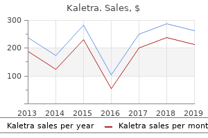
Buy cheap kaletra 250 mg on line
G treatment modalities purchase kaletra with mastercard, Higher power demonstrating mononuclear stromal cells with oval nuclei and discrete nucleoli medicine 219 buy kaletra 250 mg lowest price. Inset, An oval histiocytic cell with two nuclei and densely eosinophilic cytoplasm. Treatment and Behavior Approximately 25% of conventional giant cell tumors are considered to be locally aggressive on clinical or radiologic grounds. Curettage supplemented by cryotherapy is used in some centers to reduce the rate of local recurrence. Wide excision with allograft or prosthetic replacement significantly reduces, but does not completely eliminate, recurrences and is performed when appropriate and technically feasible. Typically, it is a result of tumor implantation into soft tissue at the time of surgical treatment. In some unusual instances, it may occur many years after the removal of the primary tumor. In the past, radiation therapy was frequently used to control the disease locally and has been proved to be effective in preventing local recurrences. Because the majority of malignant transformations in giant cell tumor are linked to prior radiation, radiotherapy is no longer recommended as a primary mode of treatment. Typically the pulmonary nodules grow slowly and are amenable to surgical excision with a prospect for cure. A, Radiograph of knee of 17-yearold skeletally mature girl with a 6-month history of knee pain whose giant cell tumor involved lateral half of tibial plateau; subchondral bone was curetted and bone grafted. D, Histologic appearance of recurrent giant cell tumor is identical to primary neoplasm. A, Radiograph of knee of a 27-year-old woman shows eccentric lytic tumor on medial side of tibial plateau. B, Eighteen months later patient returned with palpable nodule in soft tissue beneath surgical scar (arrows) with peripheral calcification seen on radiograph. E, Photomicrograph of recurrent tumor nodule with peripheral shell of reactive bone. Development of sarcoma in conventional giant cell tumor is the most serious complication but fortunately is rare. As mentioned previously, the majority of secondary sarcomas that arise in association with conventional giant cell tumor are linked to prior radiation therapy. With the decline in the use of therapeutic irradiation for giant cell tumors, malignant transformation has become exceedingly rare. Special Techniques It appears that several cell types that belong to the macrophage/osteoclastic and osteoblastic lineages contribute to the development of giant cell tumors. Ultrastructurally, the cytoplasm of mononuclear cells contains abundant rough endoplasmic reticulum, moderate numbers of mitochondria, a few lysosome-like bodies, and occasionally multiple lipid vacuoles. In summary, the ultrastructure is of little help to elucidate various dilemmas related to the origin of a giant cell tumor. It suggests, however, that the mononuclear cells have some ultrastructural similarities with cells of histiocytic lineage, macrophage lineage, or both. In fact, some of the mononuclear cells express the receptor for the immunoglobulin G crystallizable fragment and differentiation antigens associated with a macrophagemonocyte lineage. The cells of monocytemacrophage lineage do not proliferate well in vivo and are usually eliminated from tissue culture explants. In summary, the main population of cells in giant cell tumor have phenotypic features of both macrophage-like and osteoclastic cells. Gly34Trp change in the majority of cases were found in approximately 90% of giant cell tumors. Little is known about the factors governing local aggressive behavior, recurrence rate, and metastatic potential of conventional giant cell tumors. This does not correlate with the clinical behavior of the lesion and cannot be used as a reliable factor for predicting recurrence or pulmonary metastases. Allelic losses of 1p, 9q, and 19q are frequent in giant cell tumors but do not correlate with local recurrence or metastatic potential. D, High proliferation rate documented by positive immunohistochemical staining for Ki67. The key to distinguishing these lesions is in the unswerving adherence to clinicoradiologic correlation to arrive at a diagnosis.
Order kaletra 250mg without a prescription
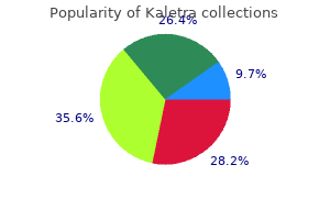
Cytologic preparations (touch smears medications j-tube cheap kaletra express, scrape medicine xyzal purchase 250mg kaletra with mastercard, Development of Bone Fetal bone formation and postnatal growth occur in one of two ways. In intramembranous ossification, clusters of fetal mesenchymal cells differentiate directly into osteoblasts. In the developing epiphyseal centers, this cartilage model undergoes focal calcification, followed by vascular invasion and the appearance of bone-synthesizing osteoblasts. The devitalized, calcified cartilage serves as a scaffolding for the deposition of bone matrix and is resorbed by osteoblasts at the same rate at which the growth plate is internally expanded. Consequently, long bone growth occurs while the thickness of the epiphyseal plate remains constant. The cessation of interstitial expansion of the epiphyseal plate results in its gradual obliteration and the termination of growth. A, Overall view of anatomy of growth plate and zone of primary spongiosa formation below. B, Zone of cartilage-cell hypertrophy at base of cartilage-cell columns where programmed cell death (apoptosis) supervenes. C, High-power view of metaphyseal side of growth plate shows osteoid deposition on surface of calcified chondroid by rimming osteoblasts. A, Low-power photomicrograph of fetal calvarial bone formation by direct osteoblastic differentiation from primitive mesenchymal cells. B, Medium-power photomicrograph shows formation of woven bone trabeculae with osteoblastic rimming without cartilage stage. More often, cytology is used as a supplement for frozen-section diagnosis, which provides an opportunity to evaluate the morphology of individual cells without freezing artifacts. Biopsy and Curettage the specimen for diagnosis can be obtained by various transcutaneous closed biopsy instruments. The use of "closed" biopsy techniques has increased, and the procedure is often assisted by various radiographic imaging techniques. These techniques are recommended as the initial diagnostic approaches, but in many cases they yield adequate material to establish the final diagnosis. However, some lesions are extremely difficult or even impossible to diagnose on the basis of a small amount of material obtained by closed biopsy techniques. Planning the open biopsy approach is a complex process that must take into account the clinical presentation, the imaging context, and the technical aspects of subsequent definitive surgery. Systematic approach with procurement of adequate samples will lead to a complete workup and more precise diagnosis. In general, the incision site and the biopsy track should be carefully selected so that if the lesion proves to be malignant, it can be excised en bloc with the segment of affected bone. Not every lesion requires microscopic verification before treatment and in some instances imaging techniques are used to guide the placement of therapeutic devices such as a radiofrequency electrode for ablation of osteoid osteoma. At the time of gross examination, a decision must be made as to whether the specimen requires decalcification or can be processed without demineralization. Routine demineralization of all bone biopsy specimens is inappropriate because this procedure destroys some cellular and extracellular components. Demineralization of the entire material may render the tissue unsuitable for some special techniques. Nucleic acids, in particular, are degraded by acid-based demineralization procedures. In general, most routine immunohistochemical studies can be performed on decalcified tissue with satisfactory results. For that reason, it is important to divide the specimen into several sections and to process the softer sections without demineralization. Occasionally, a one-step diagnostic and therapeutic procedure is chosen on the basis of the clinicoradiologic data. All tissue fragments that appear different should be submitted for microscopic examination because they may contain diagnostically important and histologically distinct components. This fixative contains glacial acetic acid in addition to picric acid, and sufficient demineralization is often achieved simultaneously with fixation. Planning of incision site so that tumor can be excised en bloc with the segment of affected bone and biopsy tract without contaminating remaining tissue.
Purchase kaletra 250mg line
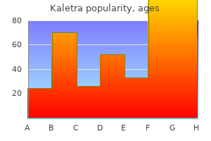
Enchondromatosis represents a pathogenetically distinct developmental disorder of enchondral ossification that is probably more accurately described as cartilage dysplasia medications for factor 8 cheap 250 mg kaletra visa. The term dyschondroplasia was introduced by Ollier in his description of the entity in 1900 medicine vs surgery purchase discount kaletra line. The lesions have a tendency to be metaphyseal and are sometimes eccentrically placed, with predominant unilateral involvement of the appendicular skeleton. The clinical manifestations typically appear during childhood, and the extent of skeletal involvement is variable. The largest series reported from the Rizzoli Institute consisted of 51 cases of multiple enchondromas presumably representing enchondromatosis compared with 334 solitary enchondromas encountered in the same period. On the other end of the spectrum are the cases with massive involvement of multiple bones and severe deformities. The most frequent presentation is a tumor affecting one extremity, but bilateral involvement is often present. Even in cases with diffuse involvement of multiple bones, the disease has a tendency to predominate on one side of the body. The bones most often affected after the hand are the short tubular bones of the feet, femur, and humerus and the bones of the forearm. The femur is the most frequently involved long tubular bone, followed by the tibia and humerus. Clinical Symptoms In general the more diffuse and severe the skeletal involvement, the earlier in life symptoms appear. In general, symptoms appear early, and most cases are typically diagnosed in childhood. Peak age incidence at onset of symptoms and most common anatomic sites of involvement. With the involvement of the long tubular bones, angular deformity of the affected extremity can be a presenting symptom. The involvement of long tubular bones is typically associated with length discrepancy. Retarded growth and length discrepancy are particularly evident in the lower extremities. In severe cases, the discrepancy can be in a range of several centimeters in early childhood (age 2 to 3 years). Pathologic fracture may be a presenting symptom, but more often occurs later in the course of the disease with the progression of bone involvement. Patients with severe enchondromatosis may be seen in adult life with shortening and bowing deformities of the extremities that severely affect motor function. Radiographic Imaging Enchondromatosis presents with radiographic features that are distinctive and in some cases diagnos- tic. Metaphyseal involvement is less evident in the short tubular bones, where the eccentricity of the lesions with multiple lytic defects oriented perpendicularly to the long axis and extending toward the soft tissue is most pronounced. The lesions often show punctate calcifications that are typical for the radiographic appearance of the cartilaginous matrix. In these locations, the lesion forms elongated grooves or longitudinal lucent columns along the long axis of bone. The radiographic appearance is best understood if the changes are envisioned as a parallel arrangement of rows of dysplastic cartilage that extend from the growth plate toward the diaphysis. With progression of the lesion as a result of the continuous growth of cartilage, larger expanding masses that extend to involve the diaphysis are formed. At this stage, the parallel arrangement of the cartilaginous lesion may become so distorted that it presents as a large multilobular mass that involves the bone end. Severe involvement of both proximal and distal metaphyses can produce a Text continued on p. Healed pathologic fracture of tibial shaft with angular deformity and bowing of fibula. Elongated columns of dysplastic cartilage extend from iliac crest growth plate into body of ilium.
Discount 250mg kaletra with amex
It provides a superior inspection of pharyngeal anatomy treatment of uti buy 250mg kaletra amex, sensation treatment zone guiseley kaletra 250 mg with visa, laryngeal closure patterns, and secretions compared with fluoroscopy. Accessibility is deemed a strength of the endoscopic procedure because of the portability of equipment and no concern of x-ray exposure posed by repeated assessments. Some clinicians and researchers have used this technique in repeated applications as a biofeedback tool, often to teach patients airway protection strategies. Perhaps the biggest limitation of the endoscopic swallowing study is the relatively limited scope of view. The image and thus evaluation focus is clearly on pharyngeal included in the endoscopic swallowing examination. Clinicians are encouraged to seek formal training in this technique because it is relatively new in the dysphagia evaluation arena and not performed routinely by all practicing clinicians. What to Look For Much like the videofluoroscopic swallowing study, interpretation of the endoscopic swallowing study also is dominated by description. In general, examining clinicians should evaluate the anatomic integrity of each "level" (velopharynx, pharynx, larynx) of the swallowing mechanism. Basic movement characteristics of each level should be documented with specific reference to absent or reduced movement. Dysphagia clinicians may also encounter additional procedures such as capsule or pill endoscopy. As the name implies, this technique requires the patient to swallow a capsule (approximately the size of a vitamin pill) containing a wireless camera that transmits pictures to a small device worn by the patient. What other endoscopic imaging techniques can you identify that may be appropriate for patients with swallowing difficulties As mentioned earlier in this section, additional research has demonstrated that neither anesthetics nor vasoconstrictors are necessary to complete this procedure. Patients who may be combative or demonstrate movement disorders that might preclude completion of a safe examination or those patients with bleeding disorders might increase any risk factor associated with this procedure. Before engaging in either the application or the interpretation of endoscopic swallowing studies, clinicians must avail themselves of an appropriate degree of supervised training (of course, this same concern should be addressed for the fluoroscopic study). Published guidelines are available from the American Speech-LanguageHearing Association that detail the knowledge and skills required to undertake this procedure and suggest mechanisms to obtain appropriate training. Some comparisons have been practical suggestions for application based on clinical experience,39,40 whereas others have been more rigorous comparisons of specific findings on the respective procedures in common groups of patients with dysphagia. Based on this advantage, the fluoroscopic procedure has been advocated as the preferred procedure for initial swallowing assessments and for imaging assessment of dysphagia symptoms focused on the esophagus. Conversely, the endoscopic procedure provides a superior inspection of anatomy and secretions. Finally, because of the portability advantage of the endoscopic procedure and the absence of radiation exposure, this procedure has been advocated for patients who are not able to be transported. Studies comparing specific findings between these two imaging procedures have consistently identified a high aspects of swallowing (see also Clinical Corner 8-3). The issue of whiteout during the swallowing peak has been raised as a potential limitation of this procedure; however, in practice this brief period of image loss rarely affects the outcome of the evaluation and, in some instances, the absence of this normal finding implicates a weakened pharyngeal swallow. Safety issues have been raised regarding this procedure; potential complications include nosebleed, laryngospasm, vasovagal response, and allergic reaction to medications when used. However, published reports of relatively large numbers of patients receiving this procedure have documented that it is a safe procedure with few complications. Thus, depending on the requirements of the clinical situation, one procedure might be indicated over the other, or the two procedures might be used in a complementary fashion during the same dysphagia evaluation. Imaging studies of swallowing provide objective imaging of the anatomy and physiology of the swallowing mechanism and swallowing biomechanics across varying bolus and patient conditions. Imaging studies of swallowing should be strongly considered whenever a thorough clinical evaluation is insufficient to answer the pertinent clinical questions for a given patient. This may include delineation of dysphagia parameters, clarification of airway protection issues, the effects of compensatory maneuvers, and monitoring changes over time. These examinations may also provide information useful in understanding medical conditions that underlie dysphagia. Commonalities exist between procedures for the fluoroscopic and endoscopic swallowing examinations. Both provide dynamic imaging of the swallowing mechanism and performance, use multiple bolus volumes and textures, and have the potential to evaluate the effect of compensatory maneuvers on swallowing safety and efficiency.
Diseases
- Cystic hygroma lethal cleft palate
- Vulvovaginitis
- Landouzy Dejerine muscular dystrophy
- Methyl mercury antenatal infection
- Fetal phenothiazine syndrome
- Thrombocytopenia Robin sequence
- Guanidinoacetate methyltransferase deficiency
- Proximal spinal muscular atrophy
- Benign astrocytoma

Kaletra 250mg free shipping
This mimicry can lead to the error of underestimating the metastasizing potential of a primary bone sarcoma when small amounts of tumor bone or osteoid formation go undetected because of sampling problems symptoms pink eye buy kaletra 250 mg low price. Careful attention to clinical and radiologic correlation helps prevent these mistakes symptoms influenza purchase kaletra 250mg overnight delivery. Chondrosarcomas are more common in patients beyond the fifth decade of life and are very rare in adolescents. Osteosarcomas with abundant cartilage matrix formation usually show radiologic features more consistent with a bone-forming tumor, including cloudlike radiodensity and prominent periosteal new bone formation in response to cortical disruption. The latter often takes the form of a sunburst or "hair-on-end" appearance that is rarely seen in chondrosarcomas. Enchondromas and low-grade conventional chondrosarcomas can be almost indistinguishable on cytologic grounds, and attention must be paid to the clinical setting and radiologic response of the surrounding bone, as well as the presence or absence of pain, the age of the patient, and whether the lesion was detected as an incidental finding. The last becomes a factor when asymptomatic enchondromas are discovered only because radiographs are obtained after an injury or bone isotope scans reveal the presence of an undiagnosed central cartilage tumor. Histologically, enchondromas tend to be less cellular, the chondroid matrix tends to be uniformly hyaline, and the matrix calcification may be abundant. The nuclei of the benign cartilage tumor cells are small, uniform, and round and have homogeneous chromatin density, in contrast to the larger nuclei with open chromatin patterns seen in chondrosarcomas. Multinucleation of chondrosarcoma cells is far more frequent than it is in enchondromas. The matrix in chondrosarcomas may show prominent myxoid change, and the tumor tends to infiltrate the intertrabecular spaces and the haversian canals rather than showing the discrete lobules bordered by lamellar bone associated with enchondromas. The difficulty in distinguishing between enchondromatosis and low-grade chondrosarcoma is further complicated by the fact that highly eccentric dyschrondroplastic lesions and even subperiosteal ones can simulate extraosseous extension of chondrosarcoma. Fibrous dysplasia with abundant cartilage differentiation (fibrocartilaginous dysplasia) may be difficult to distinguish from chondrosarcoma, particularly with limited biopsy samples having only the cartilage component. The radiographic features of fibrous dysplasia, especially when multiple skeletal sites are affected, offer a good opportunity to make this distinction. The cartilage in a lesion of fibrous dysplasia may also show microscopic evidence of an orderly enchondral ossification sequence that mimics a growth plate. Fracture callus and the exuberant reparative changes associated with pubic osteolysis can contain an abundance of proliferating cartilage tissue that sometimes 502 7 Malignant Cartilage Tumors suggests cartilage neoplasia. These can usually be recognized as nonneoplastic conditions when all of the clinical and radiographic data, in addition to the absence of true microscopic anaplasia of the reparative tissue, are considered. Synovial chondromatosis, particularly when it is associated with bone erosion, has occasionally led to the misdiagnosis of chondrosarcoma. This uncommon situation arises most often in connection with synovial chondromatosis of the hip and of the temporomandibular joint, where extension into bone by cortical erosion occurs more often. Clinical and radiologic suspicion of the synovial origin of the cartilaginous nodular tissue can often be confirmed microscopically. Conventional Chondrosarcoma in Different Anatomic Sites Similar to other bone sarcomas, conventional chondrosarcoma has identical microscopic features and biologic potential regardless of its anatomic location. However, it has a unique predilection to involve certain anatomic sites and is extremely rare in some parts of the skeleton. In general, the overall anatomic distribution of chondrosarcoma differs significantly from that of enchondroma, although some overlap exists. Moreover, the clinical significance, technical feasibility of complete removal, and chance for cure differ in relation to the anatomic site. For this reason, separate descriptions of chondrosarcoma and its differential diagnosis in various anatomic sites are provided. Approximately 40% of chondrosarcomas occur in the long tubular bones of the extremities. It is important to remember that benign intramedullary cartilage lesions of long bones are typically small and clinically asymptomatic and are often discovered as incidental findings on radiographs. Intramedullary cartilage lesions should be suspected of being grade 1 chondrosarcoma if the following features are present: foci of increased cellularity with plump nuclei and open nuclear chromatin structure; an infiltrative or destructive growth pattern; size that exceeds several centimeters in greatest dimension; pain, particularly at rest; and radiographic features of destructive growth. The pelvis is the single most frequent site involved by chondrosarcoma (~25% of all cases). The flat bones have relatively narrow medullary cavities, and cortical disruption occurs at these sites much earlier than in the long tubular bones. Virtually all chondrosarcomas of flat bones show features of cortical disruption and extensive soft tissue involvement at the time of diagnosis.
Cheap 250 mg kaletra amex
Intraoperative diagnosis is usually based on frozen sections stained with hematoxylin-eosin medications list a-z discount 250 mg kaletra fast delivery. Resection and Amputation Specimens Resection (limb-sparing procedure) or amputation is performed after the malignant or locally aggressive nature of the lesion has been confirmed by biopsy symptoms yellow fever order kaletra mastercard. Dissection of a bone resection or amputation specimen should facilitate the following objectives:51,57,58,61,68,72,85,93 1. Confirmation of the preoperative diagnosis or its modification on the basis of new information 3. The preoperative radiographs or specimen radiographs are extremely helpful in planning the dissection of resected or amputated specimens. The radiographic data assist in localizing the tumor within the specimen and identifying any areas of extension into soft tissue. They also help in the selection of the optimal plane of bone dissection and may identify special areas of interest, such as skip metastases, involvement of other structures such as a neurovascular bundle, and closest margin of resection. The best approach is to have the radiographic documentation available for review during dissection. To accomplish the goals of gross examination, it is important to perform the dissection in an organized manner: 1. Any attached soft tissue or skin should be inspected for induration, soft tissue masses, and other changes. Margins should be identified and sampled on the basis of gross examination and radiographic data. Areas of the closest soft tissue margin of excision, bone resection margin, and synovial articular resection margin should be sampled. In limb-sparing procedures, the soft tissue resection margins around the tumor are of particular importance. The potential closest margins are best identified by reviewing the clinicopathologic data in consultation with the surgeon. In long bone resection specimens, an en face proximal bone margin of excision is sufficient. Prior biopsy tracks should be left attached to bone and appropriately sampled to verify their margins and involvement by the tumor. After examination and sampling, the attached soft tissue is dissected down to the periosteum and is removed. Areas of extension into soft tissue identified on radiographs or during dissection should be left attached to the bone in continuity with the main tumor mass. Bisected specimens expose the tumor cut surface, and their gross examination can reveal features such as epiphyseal involvement, intramedullary invasion and extension into soft tissue, areas of disrupted cortex, and periosteal reaction. Pathologic assessment of the chemotherapy effect in postoperative specimens is an integral element of the multi-model approach to the management of bone tumors, and it was originally designed and clinically validated for osteosarcoma resection specimens. Sampling plan for mapping procedure of central slab to provide precise histologic information on margins of resection and extent of tumor necrosis after chemotherapy (also see Chapter 5). Theoretically the assessment of chemotherapy-related necrosis requires submission of the entire tumor for microscopic evaluation, but such an approach is impractical and dramatically increases both the workload and the technical cost of pathologic assessment. For practical purposes, it is performed from the central slice of the tumor which is subjected to specimen radiography and is subsequently divided into smaller sections submitted in individual cassettes typically averaging approximately 2. A detailed description of pathologic assessment of preoperative chemotherapy effect is provided in Chapter 5. Two staging systems are currently used to assess the clinicopathologic parameters of bone tumors. Assessment of the tumor is based on three parameters: T (tumor), N (node), and M (metastasis). T1 represents a tumor confined within the cortex, and T2 extends beyond the cortex. Nx designates the lymph nodes that cannot be assessed, N0 indicates no detectable lymph node metastases, and N1 designates the presence of metastases. Mx indicates an unknown status of metastases, M0 indicates the absence of detectable distant metastases, and M1 designates the presence of distant metastases.
Discount kaletra 250 mg line
Fibroblastic or spindle-cell meningioma is characterized by the presence of elongated spindle cells arranged in fascicles with a focal storiform pattern symptoms 0f parkinsons disease kaletra 250 mg fast delivery. Focal syncytial features with a whorled pattern and psammoma bodies are present in some fibroblastic meningiomas medicine 44334 buy kaletra 250 mg low cost. Transitional meningioma shows the presence of clearly recognizable syncytial areas and fibroblastic features. Cellular whorls are typical for this type, which is also known as mixed meningioma. Psammomatous meningioma is characterized by prominent numbers of calcifications with a concentrically layered architecture referred to as psammoma bodies. Scattered psammoma bodies can be seen in virtually any subtype of meningioma; however, they are, by definition, particularly abundant and densely packed in the psammomatous variant. The outdated term angioblastic meningioma encompassed at least three different types of tumor, including angiomatous meningioma, hemangiopericytoma, and hemangioblastoma, which are considered distinctly different neoplasms today. Metaplastic meningioma exhibits focal areas of metaplasia, which may differentiate along cartilaginous, osseous, or lipomatous lines. Secretory meningioma has microscopic features similar to those of syncytial or transitional meningioma. In addition, individual tumor cells show discrete intracytoplasmic inclusion bodies that are strongly positive for carcinoembryonic antigen. Lymphoplasmacyte rich meningioma is the least common, least well characterized, and most controversial meningioma subtype. Many examples previously reported in the literature are now recognized as reactive or inflammatory lesions, rather than meningiomas. Nevertheless, there are legitimate examples of meningiomas with prominent intratumoral lymphoplasmacytic infiltrates. Microcystic meningioma exhibits a distinctive architecture of spidery cell processes that form variously sized microcysts to the extent that, to the uninitiated, the tumor may not be recognized as a meningioma. All three of these meningioma variants-angiomatous, secretory, and microcystic-frequently display "degenerative" nuclear atypia, and in some tumors the morphologic features are so overlapping that any of the three appellations might be applied. Recognition of this entity has clinical significance because these neoplasms exhibit locally aggressive growth and are associated with late metastases. Rhabdoid meningioma is another meningioma subtype with potential for aggressive clinical behavior. Tumor cells exhibit characteristic rhabdoid morphology, often lacking other typical architectural and cytologic features of meningioma. Atypical and anaplastic meningiomas comprise the higher grade categories of meningiomas. A, Axial computed tomogram showing characteristic "hair on end" or "starburst" sign in a hyperostotic frontal meningioma. Compact fascicles of highly spindled cells (fibroblast-like) alternating with areas of syncytial appearance, giving an alternating "on-edge" and "en face" feeling. Some tumors have prominent collagen bands, which may calcify in a linear, spiculated fashion (distinct from spherical psammoma bodies). A swirling together of the syncytial and fibroblastic patterns, with resultant prominent cell whorls that constitute the hallmark feature of this subtype. Essentially a transitional meningioma in which a large percentage of the meningothelial cell whorls have calcified (formed psammoma bodies). There is no specified number, percentage, or density of psammoma bodies that is required to "qualify" as a psammomatous meningioma. As with the other grade I meningioma subtypes, there is no prognostic significance. The hallmark morphologic feature is the presence of pseudopsammoma bodies, which are brightly eosinophilic intracystoplasmic globular inclusions (proteinaceous secretions) of varying number and size. Pseudopsammoma bodies are strongly positive for carcinoembryonic antigen, and the cells that produce them are strongly positive for epithelial membrane antigen and keratin. Among the rarest of meningioma subtypes, this exhibits prominent collections of chronic inflammatory cells (lymphocytes and plasma cells).
Buy kaletra on line
A and B symptoms checklist purchase kaletra 250mg free shipping, Axial computed tomogram with contrast showing a destructive lesion involving the anterior portion of the rib treatment 5th metatarsal fracture buy discount kaletra line. C, Gross photograph of the bisected specimen shows heavily mineralized tumor involving the rib and circumferentially involving the paraosseous soft tissue. D, Low power microphotograph showing malignant bone-forming tumor with irregular patches of osteoid deposits consistent with high-grade osteosarcoma. B, Gross photograph of the resection specimen in A shows heavily mineralized tumor with destructive growth pattern and extension to the parasternal soft tissue involving the sternal manubrium consistent with osteosarcoma. E, Bisected resection specimen showing destructive tumor involving the sternal end of the clavicle with variegated mineralization pattern, cystic changes, and mucinous changes consistent with osteosarcoma. Irregular, dense lesion involves right pedicle of third lumbar vertebra, suggesting osteoblastoma. Pathologic assessment of postchemotherapy resection specimens by mapping continues to be the mainstay in evaluating tumor response to treatment and in planning alternative or further treatment in cases where there has been an unfavorable response to the initial treatment plan. The explosion of data generated by high throughput genomic approaches provides interesting insights into the complexity of molecular alterations involved in the development of osteosarcoma. There is hope that these approaches may identify novel diagnostic, prognostic, and therapeutic targets for these highly aggressive malignancies. The high level of alkaline phosphatase frequently seen in osteosarcoma is not a feature of telangiectatic osteosarcoma. Radiographic Imaging Telangiectatic osteosarcoma is typically a purely lytic lesion with a permeative destructive growth pattern. In this type of osteosarcoma, the periosteal new bone formation is often a multilayered structure referred to as onion skin. Overall, the radiographic appearance is not that of typical osteosarcoma, which has a mixture of blastic and lytic areas. This type of osteosarcoma, in particular, can have a deceptively innocent radiographic appearance with sharply demarcated margins, simulating a benign bone cyst. Gross Findings Telangiectatic osteosarcoma is very hemorrhagic, and its gross appearance is frequently described as a bag of blood. The cystic spaces can vary considerably in size and may occasionally measure several centimeters in diameter. The spongy areas represent tissue honeycombed with smaller cysts that measure up to several millimeters in size. The borders of the lesion are usually well demarcated, but often there are features of invasive growth with extensive irregular cortical erosion, complete disruption of cortical continuity, and invasion of soft tissue. With tumor progression, there is usually massive destruction of the involved bone and formation of a large palpable mass. Less frequently, instead of cystlike spaces divided by septa, there are large areas of hemorrhage with "floating" Telangiectatic Osteosarcoma Definition Telangiectatic osteosarcoma is a malignant bone-forming tumor microscopically composed of blood-filled spaces divided by septa containing neoplastic sarcomatous cells. Radiologically as well as microscopically, it superficially mimics aneurysmal bone cyst. Incidence and Location Telangiectatic osteosarcoma is rare, accounting for less than 4% of all cases of osteosarcoma. Originally, it was thought to be a variant of conventional osteosarcoma, but recently it has been categorized as a distinct form of osteosarcoma. The peak incidence and the anatomic distribution are generally similar to those of conventional osteosarcoma. It usually involves the appendicular skeleton, with more than 50% of cases located in the knee area. The distal femoral metaphysis is the single most common anatomic site followed by the proximal tibia and the proximal humerus. Approximately 10% of telangiectatic osteosarcomas are exclusively diaphyseal lesions that predominantly involve the femur, tibia, and humerus. Individual cases have been reported in flat bones, the axial skeleton, and craniofacial bones, but for practical purposes, telangiectatic osteosarcoma is a disease of long tubular bones. In rare instances, telangiectatic osteosarcoma may involve extraskeletal sites or may develop as dedifferentiation of preexisting low-grade lesions such as parosteal osteosarcoma and chondrosarcoma or other solid extraskeletal tumors. The cystic spaces have no endothelial lining, and the tumor cells are in direct contact with areas of hemorrhage. Usually there is a high degree of nuclear atypia, cellular pleomorphism, and brisk mitotic activity with numerous atypical mitoses. In addition to multinucleated sarcomatous giant cells with bizarre nuclei, there may be clearly benign osteoclast-like giant cells, which can contribute to the mimicry of aneurysmal bone cyst by telangiectatic osteosarcoma.
References:
