"Purchase 25 mg coreg otc, heart attack by one direction".
By: U. Ketil, M.A.S., M.D.
Co-Director, Center for Allied Health Nursing Education
To prove such a claim blood pressure log chart pdf order coreg 6.25mg with visa, the plaintiff must generally show that the physician or designee failed to inform the patient blood pressure examples order online coreg, parent, or guardian of a material fact relating to the proposed treatment; that consent to the treatment was given without awareness of the material fact; that a reasonably prudent patient, parent, or guardian, in a similar circumstance, would have refused to consent to the treatment if informed of such material fact; and that injuries resulted from the treatment. Like a medical malpractice claim, a failure to secure informed consent claim may succeed even if the procedure was performed without negligence or there is insufficient evidence to support a battery claim (4,17). For specific informed consents, a written or oral disclosure (in many cases, both) is required. In the case of oral disclosure, the individual obtaining consent should summarize in the patient chart, in reasonable detail, the information provided. If consent is obtained by telephone, a witness should listen to the telephone conversation and co-sign the summary in the patient chart. Disclosures about proposed procedures should be presented in terms and in a language that the patient, parents, or guardians can fully understand. At minimum, the treating physician should ensure that the following are explained: 1. Disclosures should include information about the frequency and severity of the adverse potential consequences and the likelihood, duration, and degree of anticipated benefits from the treatment(s). There is a potential to overload the consentee with information; the patient, parent, or guardian does not need to hear every possible risk, especially if the problem is extremely unlikely to occur. However, the provider should consider the disclosure carefully when there is a low risk of a problem materializing but the consequences are death or severe morbidity. The physician-centered approach measures the disclosure against the accepted practice among other physicians; it asks what a reasonable physician would disclose. At trial, the court would expect testimony of medical experts to establish the standard disclosure for a given procedure (4,18). The physiciancentered approach is problematic because the standard disclosure may not actually include sufficient information. The Specific and General Informed Consent There are two kinds of informed consent: general and specific. A patient, parent, or guardian may give a general informed consent (sometimes called a "blanket consent") when the patient is admitted to the hospital and will require ongoing clinical intervention by a number of health care providers. The patient, parents, or guardians may be kept informed of the specifics of procedures during the hospital stay, but they may not be informed of every intervention. A general informed consent will cover routine medical care that a patient may receive while in the hospital, such as drawing blood or administering a nonexperimental medication. The hospital administration will define what constitutes routine care, depending on community and professional standards. The procedures considered routine may also vary depending on the hospital unit to which a patient is admitted. Examples of nonroutine procedures might be surgery for a congenital or acquired defect, renal dialysis, and extracorporeal membrane oxygenation. In such nonroutine cases, parents or guardians must receive sufficient information specific to the procedure to permit them to make a fully informed decision. In the case of general informed consent, the patient, parent, or guardian is likely to be asked to sign a written consent; these consents should be retained with the 20 Section I Preparation and Support patient-centered approach, on the other hand, will at times require considerable time and effort, often in circumstances where time may be limited. In addition, though the patientcentered approach is meant to be based on what the parties knew before the procedure and applied objectively, in practice it can be difficult to apply so rigidly. For these reasons, courts considering informed consent issues often end up with an approach that looks more like a hybrid of the physician-based and patient-based approaches: A physician should consider what other physicians in the field would disclose in the circumstances; what this patient, parent, or guardian would want to know about the options; and his or her competence with both medical terms and with the language in use (21). There is a lot to keep in mind-including the need to consider federal or state law, hospital rules, and professional association guidelines, along with the particular circumstances of the patient and/or family. If difficult issues arise, the physician should consider consulting with hospital legal counsel before proceeding. When the patient is a neonate and, therefore, cannot express autonomy, informed consent is more complicated than when the patient is a competent adult. Coercion, Manipulation, and Persuasion Consent that is not freely given is not consent. Obviously, manipulating the parents or guardians by deliberately providing incomplete or untrue information is unacceptable. Given that there is an information imbalance between the parties, it is especially important that the information the parents or guardians receive is accurate.
A woman with infertility receives an ovary transplant from her sister who is an identical twin arrhythmia yawning purchase coreg online from canada. Agonal period is the duration between: Fundamentalsof Forensic Medicine and Toxicology 11 blood pressure 3rd trimester buy coreg amex. An old lady with mitral stenosis underwent hysterectomy for uterine fibroid and died after developing pulmonary edema. D Signs of Death 9 the changes which occur after death are helpful in estimation of the approximate time of death and to differentiate death from suspended animation. Irreversible cessation of the function of brain including brainstem: this is earliest sign of death with stoppage of functions of the nervous system. Irreversible cessation of respiration: Complete stoppage of respiration for > 4 min usually causes death. Irreversible cessation of circulation: Stoppage of heart beat for > 3-5 min is irrecoverable and results in death. Radial, brachial, femoral and carotid pulsations will be absent, if the circulation has stopped. Auscultation of heart: Absence of the heart beat over the whole precordial area and particularly over the area of the apex. Other tests: Various tests, like diaphanous, magnus, I-card, pressure, cut and heat tests are now obsolete. Later, they are constricted with the onset of rigor mortis of the constrictor muscles and evaporation of fluid. As such, their state after death is not an indication of their antemortem appearance. This haziness is transient and passes off, if a drop of water is poured on the cornea. But the cornea becomes permanently hazy after about 10-12 h of death due to decomposition. This occurs all over the body due to loss of blood pressure, but it can be seen only in retina by an ophthalmoscope. The retina is pale for the initial 2 h and the area around the disc look yellowish. Suspended Animation (Apparent Death) Definition: It is a condition in which vital signs of life (heart beat and respiration) are not detected by routine clinical methods as the functions are interrupted for some time or are reduced to a minimum. Involuntary: Seen in freezing of body, poisoning with barbiturates or opiates, newborns, drowning, electrocution, heatstroke, cholera, postanesthesia, shock, cerebral concussion or insanity. Changes in the skin and facial pallor: Skin becomes pale and ash-white due to stoppage of circulation and drainage of blood from the capillaries and the small vessels. Primary relaxation or flaccidity of the muscles: Muscles loose their tonicity and become flaccid, but the muscular tissues are still alive, their chemical reaction is alkaline and responds to electrical stimuli. Contact flattening and pallor: the areas which remain in contact with the ground, become flat and the blood from vessels of these areas is pressed out, this continues even after the formation of postmortem staining over the surrounding areas. By 12 h, the area for the disc can be known only by some convergent segmented vessels. Changes in the eye other than those in the retina and vitreous humor are less important for the purpose of estimation of time of death. The surface (outer) temperature falls more rapidly for some time than the inner core temperature. This is due to the thickness of the skin and the subcutaneous tissue which are good insulators of heat. The last part of the curve (terminal phase) is slightly above the base line which is indicative of bacterial activity.
The offence is cognizable blood pressure 220 120 effective coreg 6.25mg, bailable blood pressure medication used for hot flashes buy line coreg, non-compoundable and can be tried by any Magistrate. Indecent Assault Definition: Any unwanted sexual behavior or touching of a female without her consent, with the intention or knowledge to outrage her modesty. C Postmortem Artifacts 28 Definition: Artifacts (Latin arte: art, factum: something made) are any changes caused or features introduced in a body after death which may lead to misinterpretation of findings. Other artifacts Prinsloo Gordon artifact: A common artifact seen in all types of autopsy. It represents hemorrhage on the anterior aspect of the cervical spine, posterior to the trachea and esophagus which happens due to hypostasis. Hence, caution must be used in interpreting bleeding into the posterior neck tissues. Artifacts due to Postmortem Changes these artifacts are due to rigor mortis, postmortem staining, autolysis, putrefaction and heat. Rigor mortis: Existing rigor mortis may be broken down while removing the body from the scene of crime to the mortuary which may cause error in interpretation of time since death. Postmortem staining: Isolated patches of postmortem lividity may be mistaken for bruises. Such patches on the front and sides of the neck may be mistaken for bruising due to throttling. Pancreas is one of the first organs to undergo autolysis because of proteolytic enzymes within it, which can be mistaken for acute hemorrhagic pancreatitis. Perforation of the stomach due to autolysis have to be distinguished from that due to corrosive acid or peptic ulceration. Absence of cellular response in discolored areas establishes the postmortem origin of these changes. Specks of blood in unique and unusual areas (such as on ceilings) may mislead forensic scientists. Multiple intra-cardiac injections may result in bruising of heart and hemopericardium. It may include removal of the breasts, genital mutilation such as removal of the penis and scarification type injuries. Careful examination for violence will help in the correct diagnosis of the cause of death. Lysis of red blood cells and breakdown of vessels cause the blood to seep into the soft tissues of the scalp giving the appearance of a bruise. Skull fractures: During the opening of the skull by forceful sawing or by using a chisel and a hammer, an existing fracture of the skull may become extensive or fresh fractures may be caused. Air in blood vessels: During pulling of the dura, air may enter the blood vessels. When neck structures are pulled forcefully, air may enter the neck vessels or there may be seepage of blood around the neck structures leading to erroneous traumatic neck pathology. Visceral damage: the liver, if pulled instead of being dissected out, may cause tears in the diaphragm and laceration in the bare area of the liver.

Furthermore arrhythmia icd 9 code buy 6.25 mg coreg mastercard, several studies have shown that solid fusion and clinical outcome are not well correlated [33] blood pressure chart what your reading means buy discount coreg 6.25mg. Nevertheless, the goal must be to achieve solid fusion and it is much more likely that a poor clinical outcome and "failed surgery" with pseudarthrosis and implant failure are due to insufficient postoperative spinal stability and improper instrumentation than to excessive stability and thus stress shielding. In this context, the related question of "adjacent segment degeneration" is discussed below in detail. Occipitocervical Fixation Lateral mass and pedicular screw fixation is superior to sublaminar wiring or hooks for cervical fusions the evolution of occipitocervical fixation started with pure in-situ bone grafting, after which came wire techniques, first without and later with attached steel rods, then followed by plate/screw instrumentation in the 1990s and most recently modular combined plate-rod/screw instrumentation [46, 99, 102]. The major advantage of the latter is its greater stability, allowing the abandonment of supplemental external fixation such as halo fixators or Minerva jackets. Basically the same principles of posterior fixation as described above apply to the occipitocervical junction. Stability of occipital fixation depends on whether mono- or bicortical screws are used and the local occipital topography to the side of the screw placement. Cortical thickness is greatest at the midline and the superior and inferior nuchal lines [75]. Anterior Stabilization Principles the term "anterior instrumentation" is used for any surgical measure for the implantation of a stabilization device acting on the anterior column (according to Spinal Instrumentation Chapter 3 75 F. The surgical approach is traditionally more or less from anterior depending on the body region and the neighboring cavity. Even if in the past anterior lumbar instrumentation has been questionable for some indications in the presence of sound alternatives, in the future and with the advance of disc arthroplasty, anterior surgery will probably gain in popularity. Furthermore anterior fusion will most likely retain its position as a salvage procedure for failed disc arthroplasty. As a surgical measure interbody fusion includes an at least partial removal of the intervertebral disc and of the cartilaginous endplates and subsequent filling-up of the disc space with (structured) bone graft or nowadays increasingly with artificial spacers (cages). Nowadays a variety of cage designs are available for implantation using anterior or posterior approaches [97, 98]. While the cages retain height and provide support and stability, bony fusion occurs within and/or around the cage. However, the biomechanical requirements on these devices are very high: on one hand they should provide enough compressive strength to keep disc space height while stress concentration on the implant-bone interface must be minimized to reduce penetration or subsidence into the underlying cancellous vertebral body. On the other hand, the bone graft around and within the cage must be stressed and strained sufficiently to evoke the biological signals (release of cytokines) for bone formation [17, 84] (Table 2). In this context it is proposed that extensive stress-shielding may lead to delayed or non-union. This conflict is reflected in most current cage geometries and materials, but further work is required to fully understand the underlying mechanobiology [30]. When implanting interbody devices, the partial removal of the endplate is a prerequisite for proper graft incorporation, but a bleeding cancellous bone bed may also compromise the support of the device, especially if limited contact areas are present. Resistance to implant subsidence critically depends on the quality of underlying trabecular bone [47]. Cage designs a the first cages had a cylindrical design and were screwed into the endplates (Image Zimmer, Inc. Based on this information, an effective compromise between the biological and biomechanical requirements for fusion may be achieved by choosing larger implants with more peripheral contact areas, such as the Syncage [97]. Similar to endplate strength the overall stiffness of the stabilized spinal segment increases by a factor of three as an interbody cage is moved within the disc space towards the mechanically more advantageous anterior position [69]. The indications for anterior fusion of the spine are various and include discitis/spondylitis and vertebral burst fractures but they are still also often controversial, especially for lumbar back pain. If the surgeon decides to remove the disc, the resulting degree of instability must be estimated before choosing the type of implant and extent of surgery. It has to be emphasized that a complete discectomy combined with the dissection of the anterior longitudinal ligament renders the spine substantially unstable for all loading conditions. For flexion and lateral bending, interbody devices can restore stability profoundly. Cage kinematics Stand-alone intervertebral cages for spinal fusion exhibit poor stabilization in extension. This has led to the opinion that stand-alone cages and anterior bone grafts cause segmental distraction and thereby incongruence of the facet joints. This indicates that, with distraction of the disc space and consequent tensioned anulus fibers, a compressive force on the cage is created.
After the marrow specimen has clotted blood pressure regular cheap coreg amex, dislodge the clot gently with the use of a 1- or 2-inch needle and place it into the fixative solution prehypertension 131 coreg 12.5 mg. Process the bone marrow clot in a manner identical to a typical bone marrow biopsy, except that decalcification is not required. Chemotherapy during pregnancy and its effects on the fetus-neonatal myelosuppression: two case reports. Incidence, significance, and kinetic mechanism responsible for leukemoid reactions in patients in the neonatal intensive care unit: a prospective evaluation. Cellulitis or osteomyelitis (22) 2 these complications refer to the bone marrow biopsy procedure in general, not to the tibial site in particular. Measurement of tyrosine hydroxylase transcripts in bone marrow using biopsied tissue instead of aspirates for neuroblastoma. Normal values and examination of the blood: perinatal period, infancy, childhood, and adolescence. Infection-associated hemophagocytic syndrome due to Pseudomonas aeruginosa in preterm infants. Recommendations for the use of routine bone marrow aspirations and lumbar punctures in the follow up of patients with retinoblastoma. A comparison of iodine-123 meta-iodobenzylguanidine scintigraphy and single bone marrow aspiration biopsy in the diagnosis and follow-up of 26 children with neuroblastoma. Disposable punches ranging from 2 to 8 mm are available Specimens obtained with a 2-mm punch are very small and may not yield enough tissue for an accurate diagnosis. One recent study showed that accurate diagnoses were achieved in 79 out of 84 cases, when comparing 2-mm punch biopsies to excisional specimens (20). Skin biopsy has been performed on the fetus (11,21,22) and may be done postmortem on stillborn or recently deceased infants to produce fibroblast cultures for karyotype (see Chapter 25). Under the latter circumstances, punch or excisional biopsy from the freshest-appearing, least-macerated skin area(s) is appropriate. Genetic, enzymatic, or morphologic studies on established fibroblast strains (16) 4. Punch skin biopsy is appropriate when epidermis, dermis, and, sometimes, subcutaneous fat is required. Incisional biopsies are used predominantly for disorders of deep subcutaneous fat or fascia. Excision of larger lesions by a trained dermatologist or surgeon is preferable when planning to remove an entire large lesion. Caution should be exercised in certain anatomic locations where nerves and arteries are more superficial. Many cephalic and midline lesions may require radiologic examination prior to biopsy to rule out connection to the intracranial or intraspinal space (18,19). Avoid sites, if possible, where a small scar would potentially be cosmetically disfiguring. For suspected malignant lesions, choose more atypical areas if unable to excise completely. For large or chronic lesions, obtain specimen from periphery, including some normal skin. For most dermatoses, choose site of early or fully developed, but not end-stage, lesion. For acute eruptions and bullous disease, choose an early lesion, including some normal skin. For discrete small lesions, try to leave 1- to 2-mm margins of normal skin around the lesions. If using lidocaine with epinephrine, maximal vasoconstriction occurs at 15 minutes. Stretch skin surrounding lesion taut, perpendicular to relaxed skin tension lines.
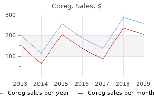
Leucoanthocyanin (Grape). Coreg.
- Are there safety concerns?
- Hayfever and seasonal nasal allergies.
- Are there any interactions with medications?
- Decreasing certain types of eye stress.
- How does Grape work?
Source: http://www.rxlist.com/script/main/art.asp?articlekey=96481
Determines the yellowness of subcutaneous tissue by measuring the difference between optical densities for light in the blue and green wavelengths b hypertension 9 code order coreg 6.25 mg without prescription. By calculating the difference between the optical densities blood pressure examples cheap 6.25 mg coreg mastercard, the parts common to the epidermis and dermis are deducted. Theoretically allows for measurement of degree of yellowness of skin and subcutaneous tissue with minimal influence of melanin pigment and skin maturity. Noninvasive device consisting of light source, microspectrophotometer, fiberoptic probe, and microprocessor control circuit b. White light is transmitted into the skin and the reflected light is collected for analysis. Algorithms take into account the effect of hemoglobin, melanin, and dermal thickness. Absorption of light due to bilirubin in the capillary bed and subcutaneous tissue is isolated by spectral subtraction. Measurements must be taken in a consistent manner with regard to placement of the probe and amount of pressure applied to the device. Interoperative and intraoperative variability may be minimized with proper training (6). Detection of hyperbilirubinaemia in jaundiced full-term neonates by eye or bilirubinometer Noninvasive measurement of total serum bilirubin in a multiracial predischarge newborn population to assess the risk of severe hyperbilirubinemia. Attempt to improve transcutaneous bilirubinometry: a double-blind study of Medick BiliMed versus Respironics Bilicheck. Skin bilirubin measurement during phototherapy in preterm and term newborn infants. Trancutaneous bilirubinometer in assessment of neonatal jaundice in Northern India. Transcutaneous bilirubin measurements and serum total bilirubin levels in Indigenous African infants. A new transcutaneous bilirubinometer, BiliChek, used in the neonatal intensive care unit and the maternity ward. Association of trancutaneous bilirubin testing in hospital with decreased readmission rate for hyperbilirubinemia. Indication Failure to locate an accessible artery or vein under normal lighting conditions for 1. Do not maintain contact between light source and extremity for long periods of time. Fiberoptic transilluminator placed on the palmar surface to visualize veins on the dorsum of hand. Optimize probe orientation, placing the target vessel in the center of the screen. Short-axis or transverse view: Probe is placed transverse to the direction of the vessel, which is seen in cross-section. Long-axis or sagittal view: Probe follows the direction of the vessel, which is seen in its length. Position the head unit at 90 degrees and approximately 13 inches (33 cm) above the target location 2. Transillumination by lightemitting diode facilitates peripheral venous cannulations in infants and small children. Prevention of burns caused by transillumination for peripheral venous access in neonates. Transilluminator burns in the neonatal intensive care unit: a mimicker of more serious disease. A cluster of atypical skin lesions in well-baby nurseries and a neonatal intensive care unit. Background the infrared light source emits a harmless, near-infrared light, which is absorbed by the blood.
In the reflectance method arrhythmia guidelines 2013 buy coreg 12.5mg without prescription, the emitter and photodetector are next to each other on the measuring site arrhythmia after heart surgery buy coreg 6.25mg low cost. Tissue composite showing dynamic as well as static components affecting light absorption. The different absorption of the wavelengths when transmitted through tissue, pulsatile blood, and nonpulsatile blood are utilized. Noninvasive continuous or intermittent arterial oxygen saturation and heart rate monitoring 2. Resuscitation Pulse oximetry is a necessary adjunct to monitoring in the delivery room. Monitoring effectiveness of bag and mask ventilation or during placement of an endotracheal tube 4. Mean arterial oxygen saturation (SaO2) values measured by pulse oximetry from the time of cord clamping. Pulse oximetry also offers an advantage for precise fraction of inspired oxygen (FiO2) control during neonatal anesthesia because of the short response time to changes in SpO2 (10). Usually, the heart rate output from the oximeter will reflect the detection of nonarterial pulsations, indicating either "0" saturation or "low-quality signal" (3). Advances in microprocessor technology have led to improved signal processing, which makes it possible to minimize motion artifact and monitor saturation more accurately during motion or low-perfusion states. Significant levels of carboxyhemoglobin or methemoglobin can yield erroneous readings (carboxyhemoglobin absorbs light at the 660-nm wavelength) (10). However, carboxyhemoglobin levels of <3% will not affect the accuracy of the instrument. Erroneous readings can occur in the presence of high fetal hemoglobin (14) A smaller effect on accuracy is noted when fetal hemoglobin levels are <50% (14). With a predominance of fetal hemoglobin, an SpO2 of >92% may be associated with hyperoxemia (14). Infants with chronic lung disease and prolonged oxygen dependence are older and have less fetal hemoglobin; therefore, SpO2 readings obtained from these 8. The same situation exists in infants who have undergone exchange transfusion because of decreased levels of fetal hemoglobin. Light sources that can affect performance include surgical lights, xenon lights, bilirubin lamps, fluorescent lights, infrared heating lamps, and direct sunlight. Although jaundice does not account for variability in pulse oximeter accuracy (15), phototherapy can interfere with accurate monitoring. Therefore, appropriate precautions should be taken, such as covering the probe with a relatively opaque material (1). Most laboratory oximeters measure fractional oxygen saturation (all hemoglobin including dysfunctional hemoglobin) as opposed to functional oxygen saturation (oxyhemoglobin and deoxyhemoglobin excluding all dysfunctional hemoglobin). Although pulse oximeters can detect hyperoxemia, it is important that type-specific alarm limits are set (16). Pulse oximeters rely on detecting pulsatile flow in body tissues; therefore, a reduction in peripheral pulsatile blood flow produced by peripheral vasoconstriction results in an inadequate signal for analysis. Pulse oximeters average their readings over several seconds depending on oximeter type and internal settings. Oximeters with a long averaging time may not be able to detect acute and transient changes in SpO2. Whenever possible, the SpO2 sensor should not be on the same extremity as the blood pressure cuff. When the cuff is inflated, the SpO2 sensor will not detect a pulse, will not update SpO2 values, and will alarm.
The principal functions of the spine are to protect the spinal cord blood pressure ranges pediatrics order 6.25 mg coreg, to provide mobility to the trunk and to transfer loads from the head and trunk to the pelvis hypertension essential discount 12.5 mg coreg with amex. By nature of a natural sagittal curvature and the relatively flexible intervertebral discs interposed between semi-rigid vertebrae, the spinal column is a compliant structure which can filter out shock and vibrations before they reach the brain. The intrinsic, passive stability of the spine is provided by the discs and surrounding ligamentous structures, and supplemented by the actions of the spinal muscles. The seven intervertebral ligaments which span each pair of adjacent vertebrae and the two synovial joints on each vertebra (facets or zygapophyseal joints) allow controlled, fully three-dimensional motion. The spine can be divided into four distinct regions: cervical, thoracic, lumbar and sacral. The cervical and lumbar spine are of greatest interest clinically, due to the substantial loading and mobility of these regions and associated high incidence of trauma and degeneration. The thoracic spine forms an integral part of the ribcage and is much less mobile due to the inherent stiffness of this structure. The sacral coccygeal region is formed by nine fused vertebrae, and articulates with the left and right ilia at the sacroiliac joints to form the pelvis. The main functions are to protect the spinal cord, provide mobility and transfer loads the spine can be divided into four distinct regions 42 Section Basic Science the Motion Segment the functional spinal unit is the smallest spine segment that exhibits the typical mechanical characteristics of the entire spine the motion segment, or functional spinal unit, comprises two adjacent vertebrae and the intervening soft tissues. With the exception of the C1 and C2 levels, each motion segment consists of an anterior structure, forming the vertebral column, and a complex set of posterior and lateral structures. The C1 (atlas) and C2 (axis) vertebrae, in contrast, have a highly specialized geometry which allows for an extremely wide range of motion at the junction of the head and neck (see Chapter 30). The neural arch, consisting of the pedicles and laminae, together with the vertebral body posterior wall form the spinal canal, a structurally significant protective structure around the spinal cord. The transverse and spinous processes provide attachment points for the skeletal muscles, while the right and left superior and inferior articular processes of the facet joints form natural kinematic constraints for the guidance of spinal intersegmental motion. Anterior Structures the Vertebral Body the trabecular bone bears the majority of the vertical compressive loads the principal biomechanical function of the vertebral body is to support the compressive loads of the spine due to body weight and muscle forces. Correspondingly, vertebral body dimensions increase from the cervical to lumbar region. The architecture of the vertebral body comprises highly porous trabecular bone, but also a fairly dense and solid shell. Vertebral body architecture and load transfer a In the healthy vertebral body, the majority of trabeculae are oriented in the principal direction of compressive loading, with horizontal trabeculae linking and reinforcing the vertical trabecular columns. The consequences are an increased tendency for individual vertical trabeculae to buckle and collapse under compressive load, as the critical load for buckling of a slender column is proportional to the cross-sectional area of the column and the stiffness of the material and inversely proportional to the square of the unsupported length of the column. Therefore, architectural remodelings which lead to a loss of horizontal connecting trabeculae are perhaps the most critical age-related changes to the vertebral body. Biomechanics of the Spine Chapter 2 Removal of the cortex decreases vertebral strength by only 10 % 43 majority of the vertical compressive loads, while the outer shell forms a reinforced structure which additionally resists torsion and shear. Previous analysis of load sharing in the vertebral body has shown that the removal of the cortex decreases vertebral strength by only 10 % [52]. However, more recent computational analyses have proposed that the cortex and trabecular core share compressive loading in an interdependent manner. The predominant orientation of individual trabeculae is vertical, in line with the principal loading direction, while adjoining horizontal trabeculae stabilize the vertical trabecular columns. Bone loss associated with aging can lead to a loss of these horizontal tie elements, which increases the effective length of the vertical structures and can facilitate the failure of individual trabeculae by buckling. The vertebral endplate forms a structural boundary between the intervertebral disc and the cancellous core of the vertebral body. With its dense cartilage layer, the endplate also serves as a semi-permeable membrane, which allows the transfer of water and solutes but prevents the loss of large proteoglycan molecules from the disc. The local material properties of the endplate demonstrate a significant spatial dependence [33]. The vertebral endplate and underlying trabecular bone together form a non-rigid system which demonstrates a significant deflection under compressive loading of up to 0.
