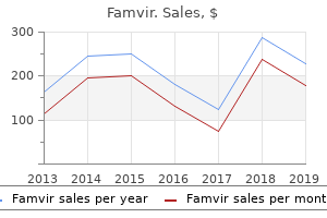
"Purchase generic famvir on line, hiv infection rates over time".
By: N. Kasim, M.A., M.D.
Deputy Director, University of Tennessee College of Medicine
Buy famvir 250mg lowest price
Most o the muscles shown move the skeleton or locomotion hiv infection detection discount famvir, but some muscles-especially those o the head- move other structures antiviral wipes purchase famvir without prescription. The sheath o the let rectus abdominis, ormed by aponeuroses o the at abdominal muscles, has been removed to reveal the muscle. Retinacula are deep ascial thickenings that tether tendons to underlying bones as they cross joints. Fusiorm muscles are spindle shaped with a round, thick belly (or bellies) and tapered ends-or example, biceps brachii. Convergent muscles arise rom a broad area and converge to orm a single tendon-or example, pectoralis major. Circular or sphincteral muscles surround a body opening or orice, constricting it when contracted-or example, orbicularis oculi (closes the eyelids). Multiheaded or multibellied muscles have more than one head o attachment or more than one contractile belly, respectively. This slight contraction, called tonic contraction or muscle tone (tonus), does not produce movement or active resistance (as phasic contraction does) but gives the muscle a certain rmness, assisting the stability o joints and the maintenance o posture, while keeping the muscle ready to respond to appropriate stimuli. Muscle tone is usually absent only when unconscious (as during deep sleep or under general anesthesia) or ater a nerve lesion resulting in paralysis. There are two main types o phasic (active) muscle contractions: (1) isotonic contractions, in which the muscle changes length in relationship to the production o movement, and (2) isometric contractions, in which muscle length remains the same-no movement occurs, but the orce (muscle tension) is increased above tonic levels to resist gravity or other antagonistic orce. The latter type o contraction is important in Equal resistance Isometric Skeletal muscles unction by contracting; they pull and never push. However, certain phenomena-such as "popping o the ears" to equalize air pressure and the musculovenous pump. When a muscle contracts and shortens, one o its attachments usually remains xed while the other (more mobile) attachment is pulled toward it, oten resulting in movement. Attachments o muscles are commonly described as the origin and insertion; the origin is usually the proximal end o the muscle, which remains xed during muscular contraction, and the insertion is usually the distal end o the muscle, which is movable. For example, when doing push-ups, the distal end o the upper limb (the hand) is xed (on the foor), and the proximal end o the limb and the trunk (o the body) are being moved. Thereore, this book usually uses the terms proximal and distal or medial and lateral when describing most muscle attachments. When studying muscle attachments, act out the action; you are more likely to learn things you have experienced. Although skeletal muscles are also reerred to as voluntary muscles, certain aspects o their activity are automatic (reexive) and thereore not voluntarily controlled. Examples are the respiratory movements o the diaphragm, controlled most o the time by refexes stimulated by the levels o oxygen and carbon dioxide in the blood (although we can willully control it within limits), and the myotatic refex, which results in movement ater a muscle stretch produced by tapping a tendon with a refex hammer. Concentric (B) and eccentric (C) contractions are isotonic contractions in which the muscle changes length: concentric contractions by shortening and eccentric contractions by actively controlled lengthening (relaxation). Muscle Tissue and Muscular System 33 maintaining upright posture and when muscles act as xators or shunt muscles as described below. The type we most commonly think o is concentric contraction, in which movement occurs as a result o the muscle shortening-or example, when liting a cup, pushing a door, or striking a blow. The ability to apply exceptional orce by means o concentric contraction oten is what distinguishes an athlete rom an amateur. The other type o isotonic contraction is eccentric contraction, in which a contracting muscle lengthens-that is, it undergoes a controlled and gradual relaxation while continually exerting a (diminishing) orce, like playing out a rope. In walking, we contract concentrically to pull our center o gravity orward, and then as it passes ahead o the limb, we contract eccentrically to prevent a lurching during the transer o weight to the other limb. Eccentric contractions require less metabolic energy at the same load but, with a maximal contraction, are capable o generating much higher tension levels than concentric contractions-as much as 50% higher (Marieb, 2016). When a motor neuron in the spinal cord is stimulated, it initiates an impulse that causes all the muscle bers supplied by that motor unit to contract simultaneously. Large motor units, in which one neuron supplies several hundred muscle bers, are in the large trunk and thigh muscles. In smaller eye and hand muscles, where precision movements are required, the motor units include only a ew muscle bers. Movement (phasic contraction) results rom the activation o an increasing number o motor units, above the level required to maintain muscle tone.
Famvir 250mg overnight delivery
The trapdoor flap requires less rotation of the coronary buttons but adds to the circumference of the proximal neoaorta hiv infection unknown order famvir 250mg overnight delivery. The intramural segment of the artery frequently passes behind the top of the posterior commissure of the original aortic valve hiv infection rate female to male order famvir 250mg with amex. The inset demonstrates that the intramural coronary artery is usually best dealt with by separating the two closely spaced coronary ostia and excising a larger than usual left coronary button often with detachment of the posterior commissure of the neopulmonary valve. This allows for less rotation of the coronary artery but increases the circumference of the proximal neoaorta, which is generally already somewhat larger than the distal divided ascending aorta. Once again, it can be seen that the coronaries are inserted above the level of the neoaortic valve and thus should not interfere in the long term with the function of the valve. The points of junction with the coronary suture lines are reinforced with mattress sutures. This commissure can be resuspended when the pulmonary artery has been reconstructed with pericardium. It is preferable not to tie the continuous suture as it passes the coronary sutures as this locks the suture line which thereby loses its ability to even out tension throughout the suture line. This is one of the great hemostatic benefits of Prolene that should not be defeated by multiple ties along its length. As an additional aid to hemostasis the three-way points of junction with the coronary suture lines are reinforced with mattress sutures. The proximal neopulmonary artery is reconstructed with a single bifurcated patch of autologous pericardium treated with 0. The patch should be larger than the combined area of the buttons excised with a view to enlarging the smaller original aorta to better match the diameter of the larger distal divided pulmonary Transposition of the Great Arteries 383 artery. The suture technique should gather the patch a little (wider bites on the patch than on the neopulmonary artery) to create slightly bulging sinuses of Valsalva. Prior to tying this suture the left heart should be filled with saline to displace air. After closing the atriotomy bypass is recommenced at a low flow rate of 50 mL/kg/min. When the posterior layer has been completed rewarming is begun and the flow rate is gradually increased. During rewarming a left atrial monitoring line is inserted through the right superior pulmonary vein. The chest is closed with interrupted stainless steel wires to the sternum with continuous Vicryl to the presternal fascia and subcutaneous and subcuticular Vicryl completing wound closure. Management of High Risk Coronary Arteries Single Coronary Artery from Right Posterior Facing Sinus with Posterior Left Main Relatively early in the arterial switch experience it was appreciated that there was a greater degree of difficulty in transferring the second most common coronary branching pattern where the circumflex coronary artery arises from the right coronary artery which itself arose from the rightward and posterior facing sinus. It was thought that this problem was primarily related to the tendency of the circumflex to kink on itself because of the acute angle resulting from transfer to the neopulmonary artery. One method for minimizing this kinking is to create a medially based trapdoor flap as described above. In addition to the kinking problem, further experience with the arterial switch elucidated another important mechanism resulting in left ventricular ischemia. From this point it branches into the anterior descending and circumflex coronary arteries and supplies essentially all of the left ventricle other than perhaps a small amount of the posterior wall if there is a very dominant right coronary system. In addition, if there has been inadequate mobilization of the left main coronary artery it may be under some degree of tension. There also may be a tendency to hinge or kink on the epicardium if the coronary artery has not been mobilized out of its epicardial fat. The initial mild degree of left ventricular global ischemia will result in left ventricular distention. In the neonate, the apex of the heart begins to point out of the chest and, in fact, may completely emerge from the chest cavity. This results in additional stress being placed on the left main coronary artery which further exacerbates either the tension or the kinking problem. Secondary myocardial edema will ensue as a consequence of the myocardial ischemia as well as the additional transfusion and higher filling pressures that are necessitated in order to maintain an adequate arterial pressure. How to Break a Positive Feedback Loop Causing Left Ventricular Ischemia the most important point is to avoid developing this feedback loop. It may be possible to mobilize an additional length of the left main coronary artery so that it is able to loop more freely rather than angling at the point where dissection from epicardial fat was stopped.
Purchase generic famvir on line
Combined Anterior and Posterior Annular Enlargement with Mechanical Valve Replacement Enlargement of the aortic annulus both anteriorly and posteriorly allows for a more symmetrical enlargement and is less likely to cause coronary artery distortion or distortion of the right ventricular outflow tract hiv infection animation 250 mg famvir visa. The procedure involves elements of both the Manougian and Konno procedures with patches placed anteriorly and posteriorly that are more modest in their width relative to what might otherwise be necessary antiviral resistance definition order discount famvir line. It is helpful to bring valve sutures from outside of the aortic wall so that pledgets lie externally. The coronary arteries must be carefully assessed as to their height above the annulus. If the coronary ostia are placed relatively low great care must be taken in using supraannular valve models, such as the St. Jude "Regent," which project above the true annulus and can impinge upon the coronary ostia. There were 20 significant complications in 16 patients: thromboembolism was noted in one patient; reoperations for aortic valve re-replacement was required in five (thrombosed valve, 3; pannus formation, 1; endocarditis 1); mitral valve replacement, 3; coronary artery bypass grafting, 2; grafting of the descending aorta, 1; and seven catheter interventions were required. At 7 years of follow-up, survival was 96% (one late death), with no differences between the infants and older patients. Freedom from mitral regurgitation mild was 100% in the older group and 41% in the infant group (p = 0. The mitral regurgitation was associated with morphologic abnormalities of the mitral valve and with development of endocardial fibroelastosis after failed intervention during the newborn period. Freedom from reoperation was 73% in the older group and 24% in the infant group (p = 0. Decision making regarding indications for and timing of surgery in these lesions is complicated. Embryology And AnAtomy Subaortic stenosis may result from simple malseptation of the original common ventricle due to poor alignment of the conal septum with the muscular interventricular septum. More than 150 mutations in 10 genes each of which encodes a single sarcomeric contractile protein have been identified. It often extends onto the ventricular aspect of the anterior leaflet of the mitral valve. Left Ventricular Outflow Tract Obstruction 433 A discrete subaortic membrane is not a congenital lesion in the true sense of the term "congenital. In these patients it has been demonstrated echocardiographically that the angle between the long axis of the left ventricle and the aorta is more acute than usual. The subsequent thickening and distortion can cause failure of leaflet coaptation or frank prolapse resulting in aortic insufficiency. Tunnel-like subaortic stenosis is often a secondary lesion that is seen following earlier resection of a simple subaortic membrane. The mitral valve also often contributes to the tunnel by being drawn anteriorly by contraction of fibrous bands which extend from the septum on to the ventricular surface of the anterior leaflet. Rare causes of subaortic stenosis include septal chordal attachments of the mitral valve as well as accessory endocardial tissue which is attached to the ventricular surface of the anterior leaflet of the mitral valve and which billows into the outflow tract during systole. Both these forms of obstruction are more common in the setting of associated heart disease such as partial atrioventricular canal defect or L-loop (congenitally corrected) transposition. However, the Venturi mechanism cannot explain the systolic anterior motion occurring at the onset of systole when outflow velocities are low. The anterior displacement of the papillary muscles produces chordal laxity in the anterior leaflet. Syncope may also occur particularly in the setting of hypertrophic obstructive cardiomyopathy. Unfortunately, sudden unheralded cardiac arrest resulting in death may be the first symptom that is encountered. It is particularly important to assess the competence of the aortic valve as new onset of aortic regurgitation is generally accepted as an indication for surgery. It can be difficult to determine by echocardiography if tissue has extended from a membrane onto the valve leaflets. Cardiac catheterization is usually not helpful in the decision making regarding need for surgery. In cases of severe concentric hypertrophy failure of coronary arterial bed neovascularization to keep pace with muscular hypertrophy will result in subendocardial ischemia and a predisposition to sudden death from ventricular fibrillation.
Purchase famvir on line amex
In patients with respiratory distress hiv rates of infection in us generic famvir 250 mg on-line, survival with homograft replacement of the central pulmonary arteries was 73 versus 41% with other techniques (p = 0 foods with antiviral properties purchase famvir 250 mg fast delivery. There were no significant differences in freedom from reintervention rates among the surgical groups (p = 0. Anomalous left coronary artery from the right pulmonary artery with aortic fusion. Cerebral metabolic recovery from deep hypothermic circulatory arrest after treatment with arginine and nitro-arginine methyl ester. Cognitive function and age at repair of transposition of the great arteries in children. Primary repair minimizing the use of conduits in neonates and infants with tetralogy or double-outlet right ventricle and anomalous coronary arteries. Anastomose zwischen System- und Lungenarterie mit Hilfe von Kunststoffprothesen bei Cyanotischen Herzvitien. Surgery of pulmonary stenosis (a case in which pulmonary valve was successfully divided). Intracardiac surgery with the aid of a mechanical pump-oxygenator system (Gibbon type): report of eight cases. Indikationsstellung und operative technik fur die korrektur der Fallotschen tetralogy. Intramural residual interventricular defects after repair of conotruncal malformations. Valved homograft replacement of aneurysmal pulmonary arteries for severely symptomatic absent pulmonary valve syndrome. Surgical treatment of absent pulmonary valve syndrome in infants: relief of bronchial obstruction. Congenital absence of the pulmonary valve: report of eight cases with review of the literature. Surgical treatment of absent pulmonary valve syndrome associated with bronchial obstruction. Pulmonary homograft monocusp reconstruction of the right ventricular outflow tract: outcomes to the intermediate term. The syndrome of absent pulmonary valve and ventricular septal defects anatomical features and embryological indications. In untreated patients with transposition and intact ventricular septum, death occurs early in infancy, generally following ductal closure at a few days of age. Not surprisingly, therefore, there were many attempts in the early years of open heart surgery in the 1950s to undertake surgical correction for these unfortunate blue babies. However, it was not until the late 1980s that anatomical correction in the form of the arterial switch procedure became the standard of care. Balloon atrial septostomy, introduced by Rashkind in Philadelphia,1 was one of the first widely applied interventional catheter techniques. Finally, children with simple transposition who have few associated extracardiac anomalies have demonstrated that it is possible to take a child with a critical, life-threatening heart anomaly to the operating room shortly after birth and to perform a major corrective open heart procedure with every expectation of an excellent outcome both in the short and longer term. The embryology of the conotruncal malformations is described in greater detail in Chapter 28, DoubleOutlet Right Ventricle. In summary, the classic theory of conotruncal malseptation suggests that failure of the septum to spiral in the usual fashion results in ventricular/great vessel discordance, that is, transposition. This results in fibrous continuity between the pulmonary and mitral valves, a hallmark of transposition. With a d-loop the ventricles lie in their usual relationship with the morphological left ventricle on the left and the morphological right ventricle anterior on the right. In contrast, levo or l-loop transposition (congenitally corrected transposition) has an entirely different pathophysiology relative to d-loop transposition (see Chapter 33, Congenitally Corrected Transposition of the Great Arteries). Embryology of thE Coronary artEriEs in D-transposition the coronary circulation develops in a similar fashion to the pulmonary arterial and pulmonary venous circulation. For example, the distal pulmonary venous system is derived from 371 372 Comprehensive Surgical Management of Congenital Heart Disease, Second Edition the original systemic venous system and invests the primitive foregut as it buds to form the primitive bronchi and, subsequently, the lungs. The original communications of the pulmonary veins with the systemic veins resorb through a system of programmed cell death similar to apoptosis. This resorption occurs when communication has been established with the primordial pulmonary vein which buds from the posterior surface of the left atrium. Failure of the pulmonary bud to link with the venous complex results in persistence of the systemic venous connection and hence total anomalous pulmonary venous connection.
Discount famvir online mastercard
Radiation treatment or pelvic cancer hiv infection per year order 250 mg famvir fast delivery, surgical complications hiv infection rate female to male purchase generic famvir on line, and infammatory bowel disease or diverticulitis may also impact the vagina. Urine enters the vagina rom vesicovaginal, ureterovaginal, and urethrovaginal stulas. Flow is continuous rom vesico- and ureterovaginal stulas but occurs only during micturition rom urethrovaginal stulas. Fecal matter or gas may be discharged rom the vagina when there is an entero- (bowel) or rectovaginal stula. Culdocentesis Manual digital examination of vagina A pelvic abscess in the recto-uterine pouch can be drained through an incision made in the posterior part o the vaginal ornix [culdocentesis-"culdo-" reerencing the term "cul-de-sac," a term used historically or the recto-uterine pouch (o Douglas)]. Medial view (from left) Pelvic Viscera 625 Laparoscopic Examination o Pelvic Viscera Visual examination o the pelvic viscera is especially useul in diagnosing many conditions aecting the pelvic viscera, such as ovarian cysts and tumors, endometriosis (the presence o unctioning endometrial tissue outside the uterus), and ectopic pregnancies. Insufation o carbon dioxide creates a pneumoperitoneum to provide space to visualize, and the pelvis is elevated so that gravity will pull the intestines into the abdomen. The uterus can be externally manipulated to acilitate visualization, or additional openings (ports) can be made to introduce other instruments or manipulation or to enable therapeutic procedures. Anesthesia or Childbirth Several options are available to women to reduce the pain and discomort experienced during childbirth. General anesthesia renders the mother unconscious; she is unaware o the labor and delivery. Clinicians monitor and regulate maternal respiration and both maternal and etal cardiac unction. Childbirth occurs passively under the control o maternal hormones with the assistance o an obstetrician. Regional anesthesia or analgesia, such as an epidural, spinal, or pudendal block, aects one area o the body. With regional analgesia, a woman is conscious o uterine contractions and can "bear down" or push to assist the contractions and expel the etus. Regional anesthesia induces complete blockade o pain and eeling and does not allow a woman to assist with labor. The anesthesia bathes the spinal nerve roots, including the pain bers rom the uterine cervix and superior vagina and the aerent bers rom the pudendal nerve. Thereore, the entire birth canal, pelvic foor, and majority o the perineum are anesthetized, but the lower limbs are not usually aected. These and the bers superior to them are not aected by the anesthetic, so the mother is aware o her uterine contractions. The perineum, pelvic foor, and birth canal are anesthetized, and motor and sensory unctions o the entire lower limbs, as well as sensation o uterine contractions, are temporarily blocked. Spinal anesthesia oten is used or limited-duration procedures, such as postpartum sterilization or orceps delivery, or or the second stage o labor. I labor is extended or the level o anesthesia is inadequate, it may be dicult or impossible to re-administer the anesthesia. Because the anesthetic agent is heavier than cerebrospinal fuid, it remains in the inerior spinal subarachnoid space while the patient is inclined. The anesthetic agent circulates into the cerebral subarachnoid space in the cranial cavity when the patient lies fat ollowing the delivery. Consequently, a severe "spinal headache" is a potential complication with spinal anesthesia that cannot occur with epidural anesthesia. With both epidural and spinal anesthesia, there is a risk that cerebrospinal fuid can leak out o the subarachnoid space. As cerebrospinal fuid leaks out, it decreases pressure within the canal, which can lead to a severe headache. It does not block pain rom the superior birth canal (uterine cervix and superior vagina), so the mother is able to eel uterine contractions. The uterine tubes are the conduits and the site o ertilization or oocytes discharged into the peritoneal cavity. Coursing in a peritoneal old (mesosalpinx) that makes up the superior margin o the broad ligament, each uterine tube has a fmbriated, unnel-like inundibulum, a wide ampulla, a narrow isthmus, and a short uterine part that traverses the uterine wall to enter the cavity.
