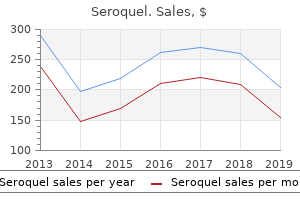
Purchase seroquel online pills
When defective ossification of the skull causes faulty development of the skull base as well medications 123 discount seroquel online american express, three common clinical features may be encountered: craniosynostosis symptoms 7 dpo bfp purchase genuine seroquel, midface hypoplasia, and exorbitism. There is greater brain injury with the presence of more premature closure of sutures, resulting in a spectrum of presentations according to the severity of anatomical and functional defects in each patient. A raised intracranial pressure may threaten survival and normal mental development may be impaired. Other natural physiological tasks such as sight, hearing, respiratory, and breathing through the nose may be compromised. Syndromic craniosynostosis, also known as craniofacial dysostosis, has sutural defects along with systemic or body involvement, for example, associated abnormalities of the limbs, spine, and heart. The array of cases ranges from extensive disfigurement reported in the medical literature to simple cleft lip and cleft palate. The etiology of craniofacial clefts is linked to abnormal development at the embryonic stage and there are various types depending on the site of origin. A 1+-year-old female patient with abnormal facial features, namely, malformed ears, down-sloping eyes, macrostomia, micrognathia, and undeveloped zygoma. There are physiological disruptions in the essential areas of airway and breathing, vision, speech, hearing, and swallowing. It has been advocated that surgery be carried out as early as possible, taking into consideration the extent of surgery and the expected blood loss. If the infant has severe protrusion of the eyes, tarsorrhaphy is recommended to sew the eyelids partially together to prevent keratitis or blindness. In a syndrome causing craniosynostosis, early screening is undertaken by a multidisciplinary team so that the comprehensive cooperative effort ensures optimum management in the planning stage, particularly when a staged surgical approach is necessary. There are a variety of surgical techniques, but meticulous planning is undertaken to choose the treatment that offers the best benefits and the least harm. Surgical procedures range from single suture correction and fronto-orbital remodeling to extensive operative techniques such as, monobloc/frontofacial advancement which is considered as early as 4 years of age. Some patients require several craniotomies and phased distractive strategies with the available tools of modern technology for better correction. To illustrate the complexity of pediatric craniofacial pathology and surgeries required, an example is given here. Cruzon Syndrome is the most frequent medical condition in syndromic craniosynostosis and is an autosomal dominant genetic disorder. Apart from significant premature suture closure, the list of systemic involvement includes maxillary hypoplasia, exorbitism, hearing loss (55%), C2C3 spinal fusion (30%), 170 L. Chan and nasal airway obstruction due to midface hypoplasia and a high-arched palate. He had congenital hydrocephalus, sleep apnea, proptosis of the eyes, bilateral hearing loss, and global developmental delay. Decision making is often not straightforward and clinical input from many professionals ensures that planning and implementation are smooth processes. Every referral of a child with craniofacial abnormality is evaluated and investigated by a group of subspecialists: pediatrician, otolaryngologist, neurosurgeon, maxillofacial surgeon, anesthetist, intensivist, plastic surgeon, geneticist, ophthalmologist, speech therapist, and psychologist. This exercise is certainly the best step to address modifiable risks to reduce morbidity and mortality. Optimization of the child can be considered the most important step for a good and safe outcome and reduces the number of "unexpected" challenges for surgery. Every clinician involved has a specific role to play and communication among the members contributes to a sound strategy to care for the child in the perioperative period. Proper feeding methods and adequate nutrition are discussed with parents so that proper weight gain is achieved. Repeated lung infections may be due to frequent aspirations from gastroesophageal reflux, esophageal dysmotility or incoordinate swallowing. Meticulous attention by the multidisciplinary team to obtain the right diagnosis assures appropriate perioperative care to prevent respiratory morbidity in the postoperative period, such as pneumonia and prolonged intubation.
Cheap seroquel 200 mg free shipping
Ineffectiveness of burst suppression therapy in mitigating perioperative cerebrovascular dysfunction medications xanax cheap 50mg seroquel mastercard. Propofol offers no advantage over isoflurane anesthesia for cerebral protection during cardiopulmonary bypass: a preliminary study of S-100beta protein levels treatment jaundice buy cheap seroquel 50mg on line. Preoperative blood glucose concentrations and postoperative outcomes after elective non-cardiac surgery: an observational study. Pharmacological experimental agents, termed blood glutamate scavengers, have been shown to improve outcomes in animal models of stroke [69], but may have unknown side effects in humans. Recently, extracorporeal methods of blood glutamate reduction, including hemofiltration, have been proposed as a safe alternative to pharmacological agents [70]. Conclusions Providing neuroprotection is the primary goal of neuroanesthesia and neurocritical care, and the discovery of therapeutic agents that meet these goals is urgently needed for patients with brain injury. To date, numerous agents have been studied across a wide spectrum of acute and chronic brain insults, many of which we have reviewed in this chapter. Although science has yet to identify a specific agent that provides neuroprotection in every context and patient population, there are several agents that have demonstrated promising results that should be investigated in future. These promising results, in combination with the devastating global impact of brain injury, justify the enormous time and costs required to move forward in the pursuit of discovering highly effective neuroprotective agents. Long-term benefits after treatment of traumatic brain injury with simvastatin in rats. Atorvastatin reduces neurological deficit and increases synaptogenesis, angiogenesis, and neuronal survival in rats subjected to traumatic brain injury. Unintentional discontinuation of statins may increase mortality after traumatic brain injury in elderly patients: a preliminary observation. N-Acetyl cysteine protects against injury in a rat model of focal cerebral ischemia. Neuroprotective effects of N-acetylcysteine on experimental closed head trauma in rats. Amelioration of acute sequelae of blast induced mild trauamatic brain injury by N-acetyl cysteine: a double-blind, placebo controlled study. Pilot study on the clinical effects of dietary supplementation with Enzogenol, a flavonoid extract of pine bark and vitamin C. The pharmacology of neurotrophic treatment with Cerebrolysin: brain protection and repair to counteract pathologies of acute and chronic neurological disorders. Cerebrolysin enhances cognitive recovery of mild traumatic brain injury patients: double-blind, placebocontrolled, randomized study. Nitric oxide modulates superoxide release and peroxynitrite formation in human blood vessels. The novel nitric oxide synthase inhibitor 4-amino-tetrahydro-L-biopterine prevents brain edema formation and intracranial hypertension following traumatic brain injury in mice. The neuroprotective effects of progesterone on traumatic brain injury: current status and future prospects. Effect of progesterone administration on prognosis of patients with diffuse axonal injury due to severe head trauma. Improved outcomes from the administration of progesterone for patients with acute severe traumatic brain injury: a randomized controlled trial. Acute alcohol intoxication and long-term outcome in patients with traumatic brain injury. Effect of elevated serum alcohol level on the outcome of severely injured patients. Treatment of increased intracranial pressure: a comparison of different hyperosmotic agents and the use of thiopental. High-dose barbiturates for refractory intracranial hypertension in children with severe traumatic brain injury. Pediatr Crit Care Med J Soc Crit Care Med World Fed Pediatr Intensive Crit Care Soc.
| Comparative prices of Seroquel |
| # | Retailer | Average price |
| 1 | Belk | 807 |
| 2 | PetSmart | 886 |
| 3 | Sherwin-Williams | 775 |
| 4 | 7-Eleven | 418 |
| 5 | Bed Bath & Beyond | 116 |
Purchase seroquel 100mg overnight delivery
Sharply incise through skin and subcutaneous fat to medicine disposal order 200mg seroquel visa, but not through medications similar to lyrica discount 100 mg seroquel overnight delivery, the quadiceps tendon epitendinous tissue. The incision through the quadriceps tendon should be 2 to 3 cm in length and full thickness, in line with its fibers. This incision should be placed 3 to 4 mm medially off midline in the event that the approach is converted to a medial parapatellar approach. Frequently, one encounters a synovial thickening that is tethering the superiormedial pole of the patella to the joint capsule. Any additional synovial folds that appear to be limiting patella excursion may also be released. If the joint continues to remain very tight, the incision should be lengthened into a medial parapatellar approach. Each manufacturer with suprapatellar instrumentation uses some variation of protection sleeve to keep patellofemoral joint cartilage uninjured during passage of reamers. Surgical Approach for Parapatellar Nailing Classically, the semiextended parapatellar approach as described by Tornetta and Collins uses a medial parapatellar incision. The patella should be evaluated before incision, and whichever side apprears to provide the most tension-free access to the entry point should be used. Regardless of which side of the patella the incision is made, the dissection remains the same. A longitudinal, 2-cm to 3-cm skin incision should be placed about the distal two thirds of the patella. Blunt dissection is carried down to retinacular tissue, which is then cleared with a sponge. The retinacular tissue is sharply incised in line with the skin incision, with care taken not to violate the synovial layer that is adhered to its undersurface. Once through retinaculum, a tissue plane is developed between synovium and retinaculum with dissecting shears. This plane is carried distally into the infrapatellar pouch behind the patella tendon placing the surgeon directly onto the tibial plateau, while remaining extraarticular. On the lateral view, the trajectory should be nearly parallel or slightly posteriorly directed (no more that 5 to 10 degrees) relative to the anterior cortex of the tibia. The first is with an awl, which typically comes on the instrument tray for the nailing system. It is provisionally malleted into the cortex, then driven in to a depth of 4 cm with a power wire driver. Once the guidewire is sunk to its final depth, its correct position should be scrutinized with fluoroscopy. This is the last opportunity to adjust the entry point before creating a large cortical entry hole. From this point and moving forward, one must ensure that the patellofemoral protection sleeve remains seated on the tibia to protect the articular cartilage. Care should be taken to meticulously guide the opening reamer through the metaphyseal bone. Changing the trajectory of the reamer is very easy, even with the guidewire in place. Fracture Reduction, Reaming, and Nail Insertion With the cortex now opened, the surgeon must pass a long ball-tip guidewire over which the nail will pass. This wire is placed into the medullary canal, across the fracture site, and seated into the distal tibia. It is not critical that the fracture be perfectly reduced to pass the ball-tip guidewire in isthmus fractures. However, in proximal or distal third fractures, where canal-to-nail diameter mismatch will occur, the fracture should be reduced before the guidewire is placed. Distally, the ball-tip guidewire (and ultimately the nail) should sit in the center of the ankle in the coronal plain, which correlates to just slightly off-center laterally in the distal tibia. Finally, once the wire is seated, it should be measured to determine the length of the ultimate nail. Many techniques exist for fracture reduction, but it should be appreciated that passage of the nail is not a reduction technique, particularly in nonisthmus fractures. Several of the reduction techniques that the surgeon should be familiar with include manual traction, strategic placement of bumps and clamps, unicortical plating, use of cortical replacing screws (also known as Poller or blocking screws), and mechanical traction through an external fixator or commercially available distractor. Failure to do so may result in eccentric reaming at the fracture site and ultimately a malreduction, which may be very difficult to overcome on passage of the nail.
Purchase seroquel with amex
Patients must lack all evidence of responsiveness treatment yeast infection men purchase seroquel with a mastercard, with absent eye opening or eye movement to noxious stimuli medications zetia discount seroquel 100 mg mastercard. There must be absence of brainstem reflexes, such as absence of pupillary response to a bright light documented in both eyes. Other motor responses that are not clear due to spinal reflexes will require an ancillary study. No eye movements should be seen in the 60 s following completion of the irrigation. Cough reflex Stimulate tracheobronchial tree by passing cannula or irrigating endotracheal tube. Hemodynamically stable without cardiac arrhythmias (Systolic blood pressure >100 mm Hg either with or without vasopressors). The apnea test should be completed as part of the first examination in which no other brain function is demonstrated. The apnea test should be completed after the motor response and brainstem reflex testing. Results of trial should be documented in medical record, including length of apneic period, blood gas results, and rate and measurable volume of breaths, if any occurred. In the above case scenario, since the cervical spine is not cleared, the oculocephalic reflex cannot be tested. Also, oculovestibular reflex pre-testing requires demonstration of patency of the external auditory canal, which may be difficult in our case. Periorbital edema may confound assessment of eye movements as well as pupillary reflex. In addition, documentation of absence of corneal reflex, absence of facial muscle movements to noxious stimuli at level of temporomandibular joints or supraorbital and supratrochlear ridges, absence of pharyngeal (gag) reflex and absence of tracheal (cough) reflex are all required. For complete description of testing for coma and brainstem reflexes, refer to Table 4. A 43-year-old male sustains motor vehicle accident with severe traumatic brain injury, and pulmonary contusions. Six hours later he is noted to be unresponsive, with dilated pupils, and no cough or gag reflex. Consideration of the pre-apnea arterial blood pH can help distinguish between these causes. A lower pH would indicate respiratory acidosis, and ventilation should be adjusted to first produce normocapnia and the test then performed. Thus, close attention must be paid to P/F ratio and systolic blood pressure prior to the initiation of apnea test. Consequently, in this clinical scenario, his hypotension would confound neurologic assessment, while PaO2/FiO2 ratio under 200 would rule out safe performance of an apnea test. A 38-year-old female is undergoing apnea testing during the process of brain death testing. The respiratory therapist places the patient on a T-piece circuit with reservoir bag, and the patient undergoes 6 min of apnea. Clinical examination demonstrated the absence of eye opening, verbal response, and motor response, as well as absence of all brain stem reflexes, with apnea on the ventilator. The treating physician is concerned for the possibility of brain death and proceeds with a formal declaration based on clinical criteria. While the patient is awaiting organ procurement, some spontaneous respirations are observed. One of the fundamental principles of assessment of patients with catastrophic brain injury is to pay close attention to the stability of vital signs.
Order seroquel 50 mg with mastercard
The evidence for this is very indirect medicine 751 cheap 50 mg seroquel fast delivery, and more studies should be conducted to assess a prognostic analysis between the different types of injuries in detail symptoms 24 hour flu generic seroquel 50 mg without prescription. Some studies have demonstrated that outcome mainly depends on the severity of the primary cerebral damage and is not worsened by the presence of extracranial injuries [22, 23]. In addition, when the motor component raises its value, better situations cannot be evaluated. On the other hand, some studies have shown that both low and high blood pressures are associated with poorer outcome [43]. However, following adjustment for age, motor score, and pupillary reactivity, the effects of higher blood pressure on outcome largely disappear, suggesting that these are merely indicative of more severe injuries and could possibly be caused by raised intracranial pressure (Cushing response). The guidelines for the surgical management of traumatic brain injury mention different cut-offs for specific traumatic lesions [52]. Various studies have found that as the extent of basal cistern compression increases from normal to partially effaced to totally effaced, mortality increases [47]. The mortality or unfavorable outcome is less for evacuated mass lesion than for nonevacuated mass lesion, though the former is mentioned as class 5, and the latter as class 6. This system has been shown to provide better prediction of outcome [49] by better discrimination Table 3. Acute pupillary dilatation in head-injured patients indicates a neurological emergency [56, 57]. All this information can improve our treatment options by characterizing functional influences, defining threshold values, and adapting therapeutic interventions in type, extent, and duration. In addition, extended neuromonitoring helps us to prevent induction of additional brain damage due to excessive therapeutic corrections. Basic neuromonitoring alone cannot assess changes in cerebral perfusion, oxygenation, metabolism, and electrophysiological function. This implies that we will miss important signs of deterioration and so we will also fail to adapt and reduce therapeutic interventions once the previous impairment has been corrected. Regarding prognosis, there are currently no genes for which the effect size is sufficiently well determined that they could be incorporated into existing prognostic models. Potential roles of genetic information may include better characterization, more accurate prognostication and therapy stratification, and identification of molecular targets for future drug development. Such knowledge might be a target for novel therapeutic interventions, drug development, and clinical trials. In the subacute phase, innate inflammatory responses decrease, while adaptive immune responses may be initiated [80]. An improved estimation of prognosis in these patients permits a more accurate clinical and ethical decision making. The exhaustive knowledge of prognostic factors offers new opportunities and should be considered an important instrument in clinical decision making and research. Relevant prognostic factors, as the ones studied in this chapter, have been identified by multivariable analysis. The injury severity score: a method for describing patients with multiple injuries and evaluating emergency care. Mortality of patients with head injury and extracranial injury treated in trauma centers. Impact of additional extracranial injuries on outcome after mild traumatic brain injury. Prognostic value of major extracranial injury in traumatic brain injury: an individual patient data meta-analysis in 39,274 patients. Predicting survival using simple clinical variables: a case study in traumatic brain injury.
Syndromes
- Smoking and alcohol use
- A personal history of colorectal cancer or polyps
- Loss of sense of taste
- Backache, which occurs with routine activities
- Transient ischemic attack (TIA), sometimes called a "mini-stroke"
- Fever
- The size, consistency, and borders of body organs
- Blood culture
- Nausea and vomiting
- Blood tests, ultrasound of your gallbladder, and other tests to make sure you are healthy enough to have surgery
50 mg seroquel with amex
Hand and finger perfusion are sometimes better indicators of vascular status than the presence of a pulse because the brachial artery may be in spasm or collaterals may provide distal circulation medications ok to take while breastfeeding buy cheap seroquel 200 mg line. The decision to surgically explore the brachial artery in the patient with a supracondylar humerus fracture with a perfused hand but no palpable radial pulse is controversial symptoms in dogs generic seroquel 50 mg on line. Certainly, pediatric supracondylar fractures with pulseless, poorly perfused limbs should undergo emergent operative reduction and fixation, and if the hand remains pulseless and poorly perfused after fixation, the artery should be explored and reconstructed if necessary. In the setting of a supracondylar fracture with a pulseless but well-perfused limb, the fracture should also undergo emergent operative reduction and fixation; however, if the limb remains pulseless but well-perfused, one can consider immediate vascular exploration or observation for 24 to 48 hours for return of pulse. The purpose of this chapter is to describe the surgical technique for closed reduction, percutaneous pinning of extension-type supracondylar humerus fractures in children. The modified Gartland system is the most common radiographic classification system for supracondylar humerus fractures and management of fractures correlates with the fracture type (Table 18-1). Other radiographic findings are used to further define supracondylar humerus fractures. Before the wide viability of fluoroscopy and familiarity with closed pinning techniques, most displaced supracondylar fractures were treated with closed reduction and splinting or casting. In the modern era, nearly all displaced supracondylar humerus fractures are treated with closed reduction and percutaneous fixation with K-wires (Video 18-1). Although most fractures may be operated on the "next day" after injury (ideally within 8 to 12 hours from injury), important exceptions that demand emergency surgery include open fractures or those that are at risk for becoming open because of skin compromise, those associated with an ipsilateral fracture in the forearm or wrist, and those with abnormal vascular examination results. Open reduction is used when acceptable reduction cannot be achieved via closed techniques, for open fractures, and for injuries associated with nerve or vascular compromise that require exploration. A short radiolucent armboard is used (as an option, some surgeons use the sterile draped C-arm platform as the operating table). Stools are placed on either side of the armboard (or C-arm) for the surgeon and first assistant. Patient Positioning the patient is positioned supine on the operating table, all the way to the side of the table. The monitor of the fluoroscopy unit is positioned so that the surgeon only has to look up from the surgical field without looking sideways or backwards. Prepping and Draping Prophylactic intravenous antibiotics are administered before prepping. Alternatively, semisterile technique may be used because results are similar; the limb is prepped sterilely and the surgeon and assistant wear sterile gloves, but gowns and drapes of the entire field are not used. Reduction Apply longitudinal traction with the elbow flexed to about 20 to 30 degrees while an assistant provides countertraction against the upper arm. The "milking maneuver" may be used if the proximal fragment has pierced the brachialis. Correct malrotation by securing the humeral shaft while rotating the forearm and distal fracture fragment together. If the fracture cannot be reduced and a rubbery or no bone-on-bone feeling is experienced, the fracture likely needs an open reduction to remove the soft tissue or potentially the neurovascular structures (median nerve or brachial artery) from the fracture site. Excessive manipulation greater than two to three attempts at closed reduction can lead to excessive swelling and increase the risk for development of a compartment syndrome. The lateral view is obtained by rotating the humeral shaft, not the forearm, to ensure that reduction is not lost. Up to approximately 25% to 30% of translation of the distal fragment is acceptable in either plane, as is a mild-moderate amount of rotational malalignment. Pin Placement Position the elbow on a towel and palpate the lateral humeral condyle. If correctly positioned, the K-wire is placed through the skin into the cartilage of the distal lateral condyle, and the start point and trajectory are confirmed with fluoroscopy. The wire is advanced with a drill, engaging the distal fragment and then the medial cortex of the humerus proximal to the fracture site. The feel of the wire advancing through the proximal cortex indicates that the wire has bicortical purchase, a key to stable fixation. The pin may cross the olecranon fossa to increase the number of cortices captured by the pin, thus improving fixation, but the arm is not fully able to extend until the pin is removed. In the coronal plane, the ideal pin configuration is one in which all wires are divergent and maximally spread across the fracture site, with at least one pin bridging the medial column of the distal humerus.
Buy seroquel on line
The modified forceps have divergent branches that are shorter and easier to handle than the conventional version [6] treatment 8th feb buy seroquel 300mg otc. Once the forceps are applied atlas genius - symptoms generic 50 mg seroquel, flexion of the head is achieved by delicately lifting the legs and lowering the fetal head toward them. Vacuum extractor the use of an obstetric ventouse for fetal head extraction during a cesarean delivery was described for the first time by Solomons in 1962 and is an excellent alternative to the use of forceps [7]. After the uterine incision, the assistant generally stabilizes the head on the lower uterine breach and exerts a pressure on the uterine fundus. Later, numerous soft and semirigid cups were manufactured, which contributed to the increase in use of the obstetric ventouse. Indeed, starting in the 1970s, the obstetric ventouse was the most widely used instrument in vaginal deliveries [9]. In certain cases, the new "soft" obstetric ventouses that improve the extraction of the fetal head are used even during cesarean delivery. An example is the "Kiwi" single-use ventouses of which there are two types: the OmniCup and the ProCup [10]. The Kiwi OmniCup is suited for all fetal head positions including posterior asynclitism and lateral malposition. Traction can be regulated even in case of contamination of the cup with amniotic fluid or blood. The obstetrician pulls on the fetal head in an upward direction so that the chin of the fetus can emerge from the the presented part without detaching it from the pulling instrument. Unfortunately, the presented part is frequently malpositioned, especially in case of asynclitism and deflection. In cases such as these, the Kiwi OmniCup is practical, flexible, and does not cause trauma. It has thus proven to be better than traditional ventouses and can also be used for transverse and occiput posterior positions. This is especially true for a cesarean delivery in which the cup should be applied on any part of the scalp, except on the face and ears. Literature contains comparative studies and meta-analyses on the application of both rigid and soft ventouses during vaginal delivery. There are, however, few references on the application of these instruments during a cesarean delivery [11]. Compared to vaginal delivery, soft ventouses reduce the risk of damage to the fetal scalp. However, it does not seem to reduce the more serious fetal lesions, such as subaponeurotic and intracranial hemorrhages. In addition, when applied outside the occiput, it has a higher risk of failure [12]. It seems therefore reasonable during a cesarean delivery to use soft ventouses for extractions in which the position of the fetal head is not especially difficult and in which a pulling force is sufficient. To correctly apply the "soft" vacuum, once the lower uterine segment has been cut, start out by locating the fetal occiput so that the cup can be correctly applied on the fetal scalp. The use of forceps or obstetric ventouse in a cesarean delivery depends on the experience of the operator and whether special cases are present, such as fetal malformations [13]. The forceps present a risk of facial and intracranial damages, whereas the risks posed by obstetric ventouses are not as severe. The pressure needed to create a vacuum on the fetal scalp is applied on the Kiwi OmniCup and is shown on a scale bar inserted in the manual pump: clinical studies recommend a pressure between 450 and 600 mm Hg (green zone) and in particular below 620 mm Hg (red zone). In particular, fetal damage of the dura mater results from repeated applications of the vacuum during particularly difficult extractions. Conclusions the cesarean delivery is an intervention that has been created to facilitate abdominal extraction of the fetus, which would otherwise be difficult or impossible through vaginal delivery. Generally, the extraction is carried out manually by an operator who may be assisted by a "Kristeller" applied on the uterine fundus by the assistant. However, in certain cases forceps and ventouses may prove useful in the extraction of the fetal head from the hysterotomy. Currently, a ventouse is preferred to forceps due to the fewer numbers of complications, especially to the fetus, that arise during their use [15].
Cheap seroquel on line
Cardiologists will often see this on an Cardiovascular Hemodynamics for the Clinician treatment concussion buy cheap seroquel on line, Second Edition moroccanoil treatment discount 300 mg seroquel overnight delivery. This is felt secondary to hypoxiatriggered vasoconstriction and vascular remodeling, as well as hyperin flationinduced compression and obliteration of alveolar vessels. This collection of disorders has unclear and/or multifactorial mechanisms as to the causation of pulmonary hypertension (see Table 26. Pulmonary artery pressures increase in response to elevated left atrial pressure, cardiac output, or true changes in pulmonary vascular resistance from arterial remodeling. Thus, hemodynamics obtained through right heart catheterization are imperative to guide diagnosis and treatment options. Hemodynamic changes in patients with elevated pulmonary pressures depend on the extent of the pulmonary hypertension. The sheer magnitude to which the pulmonary pressures are elevated is important to note; the higher the pressure, the more advanced the disease. The thinwalled, highly compliant right ventricle can accommodate acute increases in volumes at physiologic pressures. However, over time, as pulmonary vascular resistance increases with obliterative pulmonary artery remodeling, the right ventricle reaches a point where it can no longer overcome this afterload. As right ventricular failure ensues and forward flow is impaired, the mean pulmonary artery pressure will decrease. This endstage disease progression is best detected by significant elevations in right atrial pressures (generally >20 mm Hg) associated with a low cardiac index (<2. In more severe cases with significant tricuspid regurgitation, promi nent V waves may be present on right atrial pressure tracings. Assessing pulmonary vasoreactivity with pharmacologic vasodilator therapy is indicated in several instances. This test predicts response to oral calciumchannel blocker therapy and has the most evidence for treatment effect among the etiologic groups listed earlier. This is most easily accomplished with inhaled nitric oxide, although Pulmonary hypertension 315 occasionally intravenous adenosine or epoprostenol is employed. This results from longstanding, untreated large intra and extracardiac defects that initially begin as systemic to pulmonary shunts. It is felt that highpressure pulmonary blood flow results in vascular endothelial damage that incites the milieu to shift into the remodeling process of vasoconstric tion, inflammation, and thrombosis. It remains important to distinguish these components, because donor hearts are unable to pump against significant pulmonary resistance for any length of time. If there is a large component of fixed resistance, the cardiac allograft will fail, because the donor right ventricle cannot acclimate and overcome the elevated afterload. Peak systolic and mean pressures of 120 mm Hg and 82 mm Hg are indicative of severe pulmonary hypertension. Panel (c) shows simultaneous measurements of right and left ventricular pressures. Nitroprusside is the preferred agent of choice, since it induces a simultaneous decrease in both Pulmonary hypertension 317 pulmonary and systemic afterload. Inability to "reverse" pulmonary hypertension is associated with a high incidence of posttransplant mortality from right heart failure, and is thus a contraindication to heart transplantation. This results from increased right ventricular myo cardial oxygen demand due to high wall stress. There can also occasionally be compression of the left main coronary by a dilated main pulmonary artery. On physical exam, increased 318 Cardiovascular hemodynamics for the clinician jugular venous pressure with visually prominent A waves is typical. A left parasternal lift produced by the impulse of the hypertrophied highpressure right ventricle is sometimes palpable. On auscultation, patients with pulmonary hypertension will have an increased pulmonic component of the second heart sound and may have a rightsided S4 heart sound and/or an early systolic ejection click due to sudden interruption of pulmonary valve opening.
Safe 100 mg seroquel
Neuroprotective effects of erythropoietin in patients with severe closed brain injury medicine balls for sale purchase seroquel 50mg mastercard. Effect of erythropoietin and transfusion threshold on neurological recovery after traumatic brain injury: a randomized clinical trial treatment 4 ringworm buy seroquel 50mg free shipping. Republished: Society for Neuroscience in Anesthesiology and Critical Care expert consensus statement: anesthetic management of endovascular treatment for acute ischemic stroke. Type of anesthesia and differences in clinical outcome after intra-arterial treatment for ischemic stroke. Response to letter regarding article, "type of anesthesia and differences in clinical outcome after intra-arterial treatment for ischemic stroke". Extracorporeal methods of blood glutamate scavenging: a novel therapeutic modality. With the development of new knowledge about germs in the nineteenth century, sepsis was reconsidered as a systemic infection, i. Now even this concept has been further revised, since we know that infection per se is necessary but not sufficient to generate sepsis, being this the result of the interaction between microorganism and host reaction. Another important aspect is that sepsis must be viewed in the majority of cases as one step of a complex and multiphase pathological process that can be elicited even by a noninfectious cause. Therefore, an estimate of more than 750,000 cases per year in the United States and 19 million all over the world can be considered reasonable, albeit probably underestimated [2, 3]. With respect to the kind of patient more exposed to the risk of developing sepsis, we observe from epidemiological data that there is a peak of incidence in the first year of life, a relatively low incidence in adolescents and young adults, then cases become progressively more frequent around the early 50s, with the highest incidence between 55 and 65 years of age, with a slight prevalence of males over females and of blacks over whites [4]. Hospitalized patients are far more at risk for sepsis than ambulatory ones, especially after trauma or surgical procedure, or when immunosuppression of any cause and debilitating chronic disease is present. Tachycardia is an important sign, but must be viewed taking into account rhythm pattern and underlying cardiac disease, together with the use of drugs potentially interfering with heart rate, while an increased respiratory rate is a key and sometimes early warning. Besides tachypnea, typical of all patients, hypoxemia is present in the vast majority of them (PaO2/FiO2 < 300), so that oxygen administration and mechanical ventilation are, respectively, always and very often necessary. The typical clinical scenario of severe sepsis is a patient with respiratory and circulatory failure; then, it is very common for hypotensive patients to develop oliguria or overt renal failure in cases of persistent shock or use of nephrotoxic drugs. During the subsequent evolution of severe sepsis, coagulation abnormalities are found very often, ranging from low platelet count to intravascular disseminated coagulation, as well as signs of liver, brain, and bowel failure. Alveolar filling with inflammatory exudates and capillary clotting are only two of the main pathological findings; the functional result is worsened oxygenation, increased airway resistance, and decreased compliance with overall increased respiratory load. The coexistence of hypotension and circulatory failure decreases blood flow to the respiratory muscles with consequent ventilatory pump failure and need for artificial respiratory support. Besides conventional diagnostic tools for the diagnosis and monitoring of lung involvement (chest X ray, oxymetry and arterial blood gas analysis), the use of lung ultrasound is at present a well-recognized and reliable noninvasive tool [8]. The cardiovascular system is involved in the majority of patients suffering from severe sepsis and in all patients with septic shock. The typical circulatory profile includes hypotension, reduced central venous and capillary wedge pressure, and these are generally the consequences of increased vascular permeability with interstitial fluid shift, together with reduced intake and increased loss (sweating, fever, tachypnea, vomiting etc. Cardiac output is typically normal or increased together with low systemic vascular resistance; however, due to the possible myocardial depressant effect of circulating toxins, it is not uncommon to find a picture with low cardiac output and high filling pressure, or simply a flattened cardiac performance/filling pressure slope after optimal fluid replacement, indicating limited cardiac reserve. Ceriana hours and choice of optimal vasopressors, generally norepinephrine for maintenance of systemic vascular resistance and tissue perfusion and dobutamine when an inotropic agent is required. In order to maintain adequate oxygen delivery and to avoid excessive hemodilution, transfusion of packed red blood cells must be considered in order to maintain hemoglobin concentration in the range between 7 and 9 g/dl. Renal function during severe sepsis shows a variable degree of impairment; oliguria and rise of blood creatinine up to 2. The protective use of low-dose dopamine lacks strong clinical evidence and is rarely used. The avoidance of nephrotoxic agents and drugs is mandatory, together with the maintenance of adequate blood volume and perfusion pressure. Renal replacement therapy can be applied through different techniques and is strictly necessary only in a minority of cases and for a limited period of time, with restoration of normal kidney function in almost all survivors, even if this is strongly influenced by previous renal function, being patients with diabetes and multisite atherosclerotic vascular disease at increased risk to need long-term dialysis.
100mg seroquel with visa
Clinical surrogates treatment 197 107 blood pressure purchase seroquel 100 mg free shipping, such as blood pressure medications rheumatoid arthritis discount 50mg seroquel with amex, pulse rate, central venous pressure, and arterial oxygen saturation have not consistently proven useful in guiding use of therapies to maximize cardiac output. In the absence of a measurement of cardiac output, volume resuscitation is often the first approach to treating shock. However, studies have shown that tissue perfusion does not increase in response to volume resuscitation in a significant number of hemodynamically unstable patients. In the nonresponsive patients, volume resuscitation does not improve stroke volume and carries some risk of harm. Therefore, there is a large interest in developing methods to measure cardiac output continuously and accurately in critically ill patients. This would enable accurate fluid administration and optimal use of inotropic agents and vasopressors to maximize tissue perfusion [14]. Traditionally, continuous cardiac output monitoring has been accomplished using a pulmonary artery catheter. An invasive method that is useful only in ventilated patients involves using an esophageal Doppler probe to measure velocity and diameter continuously in the descending aorta. Bioimpedance systems measure the resistance to flow of electric current through the thorax. This resistance is referred to as impedance (Zo), which in the thorax includes impedance due to tissues that do not change over time (such as lung, muscle, bone, and fat) and impedance due to the amount of blood in the thorax. Thus Zo varies in proportion to the amount of fluid in the thorax and the instantaneous rate of the change of Zo is related to instantaneous blood flow in the aorta. The limitations of bioimpedance systems are many and include being sensitive to the placement of the electrodes on the body, variations in body size, and other physical factors such as temperature and humidity that have an impact on electric conductivity. Newer technologies have been developed, including bioreactance, which is used to measure the phase shift in voltage across the thorax. Pulsatile ejection of blood leads to instantaneous changes in the phase of Zo; phase shifts in Zo can thus be used as a surrogate for aortic flow. Assumed oxygen consumption based on calculation from dye dilution cardiac output: an improved formula. Use of assumed versus measured oxygen consumption for the determination of cardiac output using the Fick principle. Comparison of thermodilution and Fick methods for measurement of cardiac output in tricuspid regurgitation. Underestimation of cardiac output by thermodilution in patients with tricuspid regurgitation. Cold vs room temperature injectate and the importance of measuring the injectate temperature in the right atrium. Stouffer In the normal circulation, blood passes from the venous system through the right heart, into the pulmonary circulation, then into the left heart, and finally into the systemic circulation in a continuous, unidirectional manner. More rarely, unoxygenated blood can be shunted from the right heart to the left heart. Intracardiac shunting of blood results when there is an opening between the right and left heart chambers and a pressure difference to drive flow between the connected chambers. We will not address these shunts in this chapter, but rather concentrate on intracardiac shunts. Detection of an intracardiac shunt the presence of a shunt can be determined either invasively or noninvasively. The indicator dilution method is of historical interest, but is rarely used outside of research studies. It involves injecting a substance, such as indocyanine green, into the venous systems or a right heart chamber and then monitoring its appearance in the systemic circulation. Dye curve measurements are very accurate, but are slow and require Intracardiac shunts 93 specialized equipment that is rarely available in modern catheterization laboratories. Contrast angiography, in which contrast dye is injected into the higher pressure chamber of a suspected shunt.
References:
